HOME > Weekly Case > Weekly Case
Weekly Case
종결
Case No. 501
2018-12-01~2018-12-31
출제자 : 유인선
Hit : 532
| Period | 2018-12-01~2018-12-31 |
|---|---|
| Diagnosis | Langerhans Cell Histiocytosis (LCH) |
| Gender |
|
| Age | 64 |
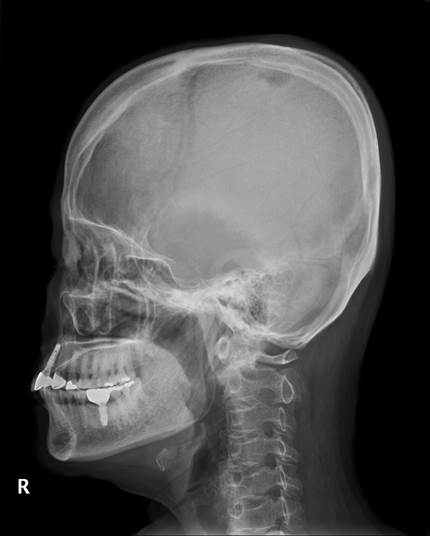
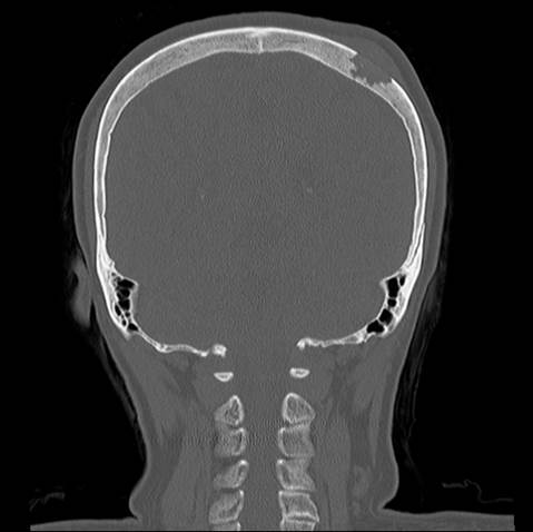
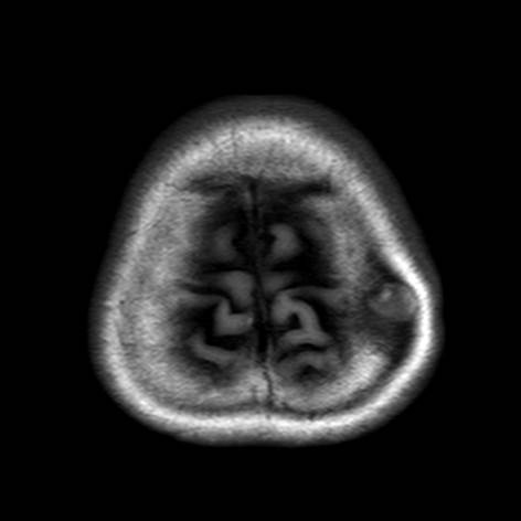
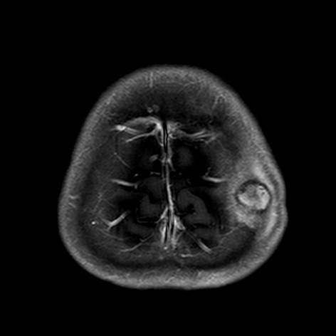
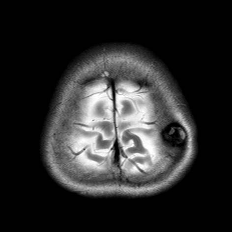
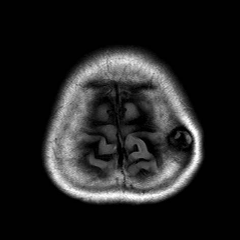
| Clinical information | F/64 Palpable lesion in scalp, left parietal area (O: 1MA) |
| Discussion | Langerhans Cell Histiocytosis (LCH) •Clonal proliferative disorder of cells of the mononuclear phagocytic and dendritic cell system that often presents in childhood 1. solitary often curable bone lesion 2. widespread, often multi-system, sometimes lethal disorder •Age: from the newborn period to 15 years of age, with a peak incidence btw 1 and 4 years of age, male predilection (2:1) •3 groups based on number of lesions and systems involved 1. The unifocal (localized) form; 70% of LCH cases, is limited to a single bone or a few bones, and may involve the lung. 2. The multifocal unisystem (chronic recurring) form; 20%, involves multiple bones as well as the reticuloendothelial system, often is accompanied by diabetes insipidus. 3. The multifocal multisystem (fulminant) form; 10%, often fatal, disseminated involvement of the reticuloendothelial system, anemia, and thrombocytopenia • Imaging findings of Bone lesion 1. Bone lesions are the m/c radiographic manifestation of LCH (~80%) 2. has a predilection for the flat bones (m/c skull) 3. The well-defined lytic “punched-out” appearance of skull lesions 4. Beveled edge - caused by asymmetric destruction of the inner and outer cortices 5. Lacks periosteal reaction in skull (unlike other osseous involvement) 6. T1 iso, T2 hyperintense, enhancing inner soft tissue component. |
| Correct answer |
Correct Answer
Semi-Correct Answer |