HOME > Weekly Case > Weekly Case
Weekly Case
종결
Case No. 500
2018-12-01~2018-12-31
출제자 : 유인선
Hit : 330
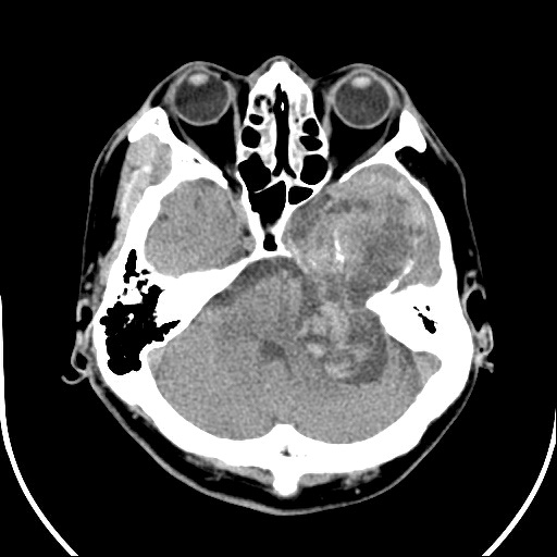
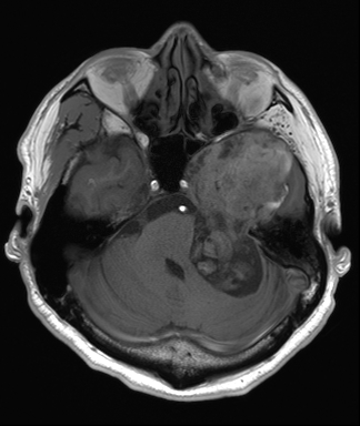
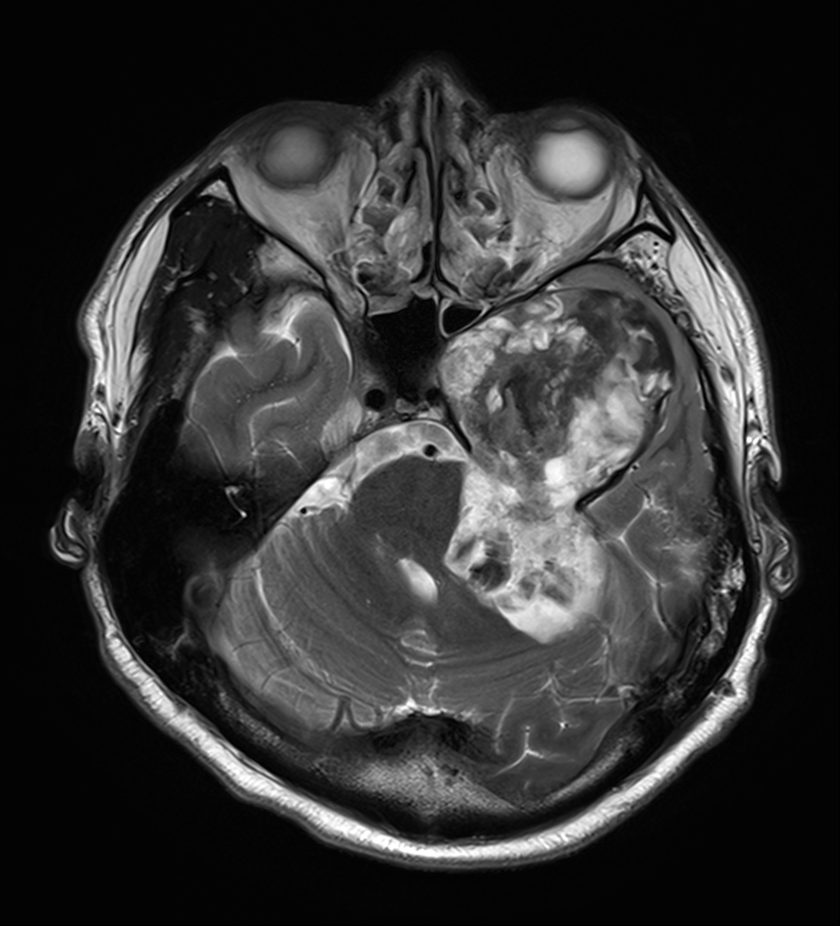
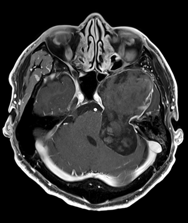
| Period | 2018-12-01~2018-12-31 |
|---|---|
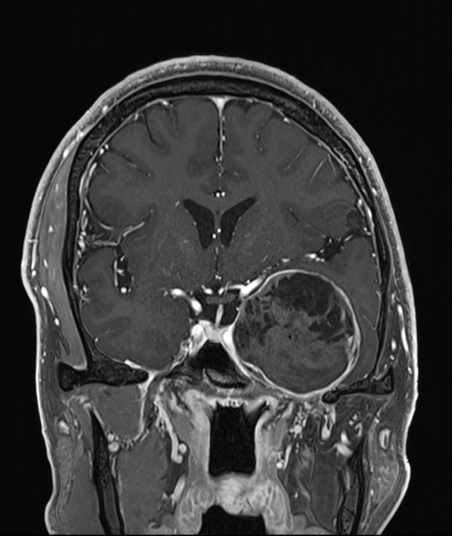
| Diagnosis | Intracranial Epidermoid cyst |
| Gender |
|
| Age | 51 |
| Clinical information | M/51 30년전부터 시작된 좌측 청력저하, 얼굴감각 저하 |
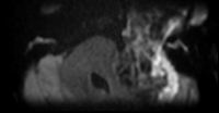
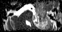
| Discussion | Intracranial Epidermoid cyst Primary intracranial cystic lesion, 1% of all cranial tumors Congenital: Inclusion of ectodermal elements during neural tube closure Acquired: Postsurgical, post-traumatic Epidemiology: 20-40 years of age, M=F Symptoms: headache, CN deficits, cerebellar sx, seizures Location : Cerebellopontine angle (~50%), 4th ventricle, sellar/parasellar Imaging findings CT Low density due to cellular debris, high cholesterol, resemble CSF Calcification (10-25%) Enhancement (-), Peripheral enhance (25%) MR Isointense to CSF (T1 hypo, T2 hyper SI) FLAIR usually doesn’t completely null DWI : Diffusion restriction Water molecules linked to cholesterol molecules in the cyst keratinaceous debris within the lesion Treatment Surgical excision if symptomatic Recurrence common |
| Correct answer |
Correct Answer
Semi-Correct Answer |