HOME > Weekly Case > Weekly Case
Weekly Case
종결
Case No. 493
2018-09-01~2018-09-30
출제자 : 박미나
Hit : 299
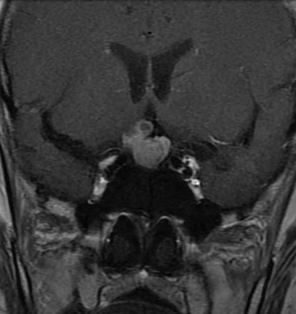
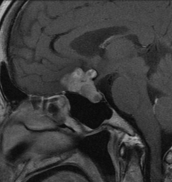
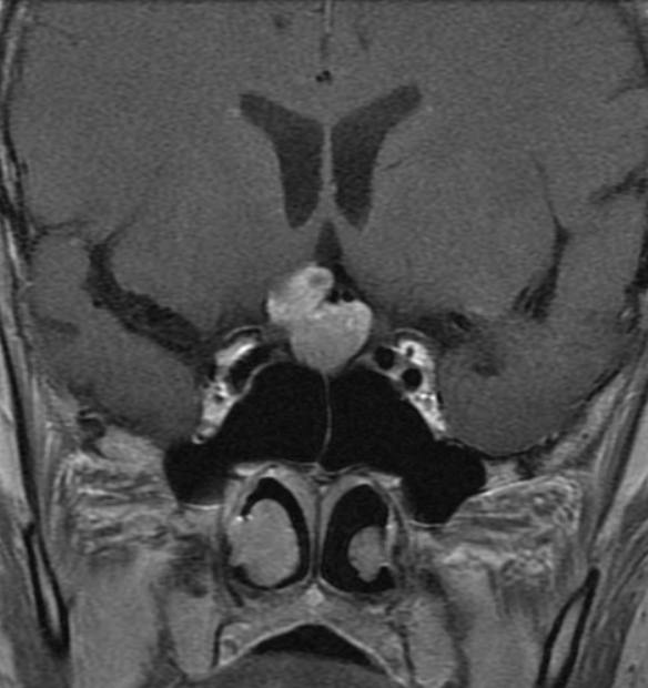
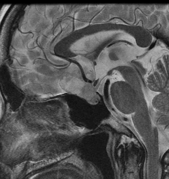
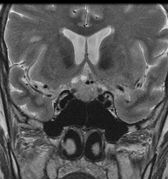
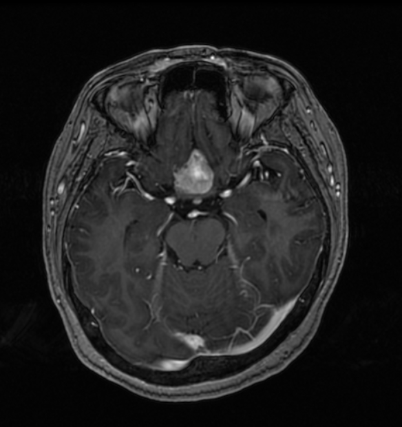
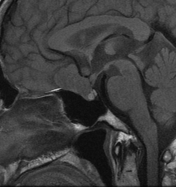
| Period | 2018-09-01~2018-09-30 |
|---|---|
| Diagnosis | Intrasellar paraganglioma |
| Clinical information | 60세 여환 우연히 발견된 뇌하수체 종양으로 내원. |
| Discussion | Background: Paragangliomas are infrequent neuroendocrine tumors of neural crest origin and they are usually benign and occur in diverse anatomic sites of the body. Paragangliomas in the sellar region are extremely rare, accounting for 0.012% of cervicocephalic tumors. There is no definite explanation for the development of paragangliomas at sites and the hypothesis is that they result from aberrant migration of small aggregates of paraganglion cells along the tympanic or ciliary nerves or branches of the glossopharyngeal nerve within or close to the cavernous sinus. Key Diagnostic Feature: The MRI features of published cases are homogeneous masses, low signal intensity on T1WIs and high signal intensity on T2WIs. Following the intravenous administration of contrast material, there was a homogeneous pattern of enhancement. The classic “salt and pepper” appearance of paraganglioma of head and neck is not expected in a sellar location The imaging pattern of paragangliomas is not specific and can mimic a meningioma or a pituitary adenoma. |
| Correct answer |
Correct AnswerSemi-Correct Answer |