HOME > Weekly Case > Weekly Case
Weekly Case
종결
Case No. 492
2018-09-01~2018-09-30
출제자 : 박미나
Hit : 294

| Period | 2018-09-01~2018-09-30 |
|---|---|
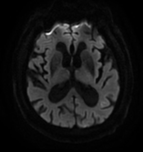
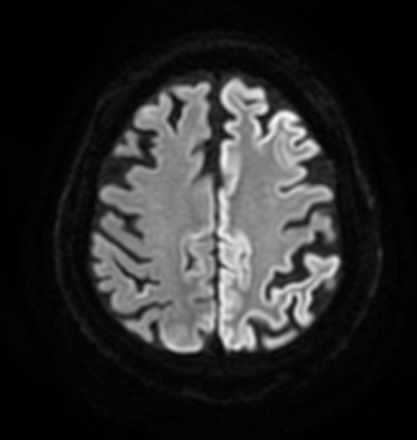
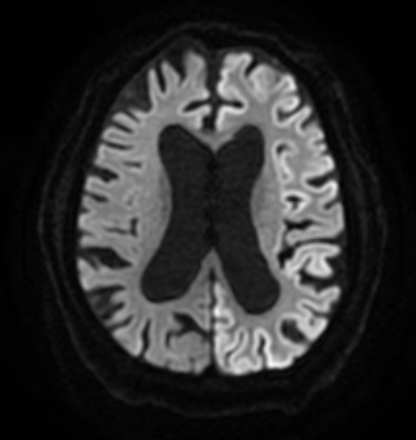
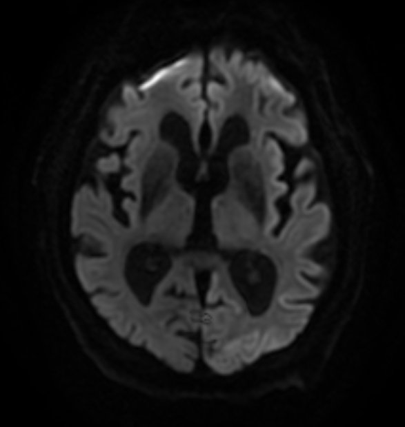
| Diagnosis | Spontaneous Creutzfeldt–Jakob disease |
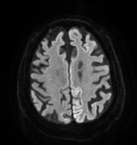
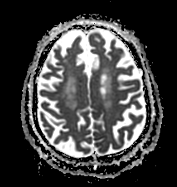
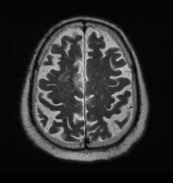
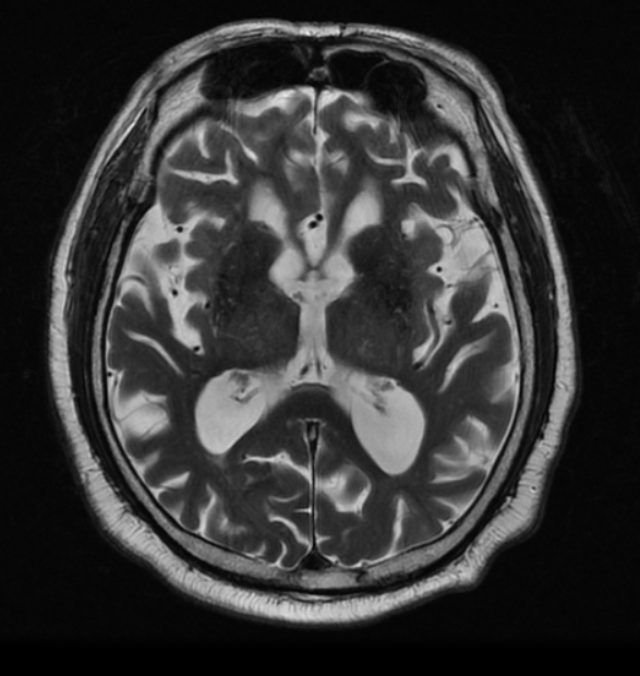
| Clinical information | 87세 남환 2개월전부터 빠르게 진행되는 인지기능 저하로 내원 그림7-9는 2개월후 FU DWI영상임. |
| Discussion | Background: Spontaneous Creutzfeldt–Jakob disease (sCJD) is a uniformly fatal neurodegenerative disorder caused by the accumulation of an abnormal form of the human prion protein PrPSc in the brain. Clinical diagnosis is based on the combination of rapidly progressive dementia, myoclonus and multifocal neurological dysfunction associated with an electroencephalogram showing generalized periodic sharp wave complexes and/or a positive 14-3-3 protein test in CSF. Clinical Presentation: CJD is a progressive, fatal illness. Over 90% of patients rapidly progress from normal cognitive function to death in under a year. Key Diagnostic Feature: MR with DWI is the imaging procedure of choice. T1 scans are often normal but may show faint hyperintensities in the posterior thalami. FLAIR hyperintensity or restricted diffusion in the caudate nucleus and putamen or in at least two cortical regions (temporal-parietal-occipital "cortical ribboning") are considered highly sensitive and specific for the diagnosis of sCJD. Differential Diagnoses: CJD must be distinguished from other causes of rapidly progressive dementia, such as viral encephalitis, paraneoplastic limbic encephalitis, and the recently characterized autoimmune-mediated inflammatory disorders, such as voltage-gated K-channel, NMDAR, or GABA encephalopathies. |
| Correct answer |
Correct Answer
Semi-Correct Answer |