HOME > Weekly Case > Weekly Case
Weekly Case
종결
Case No. 472
2018-02-01~2018-02-28
출제자 : 배윤정
Hit : 313
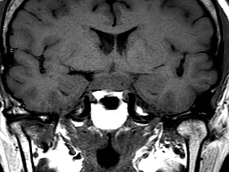
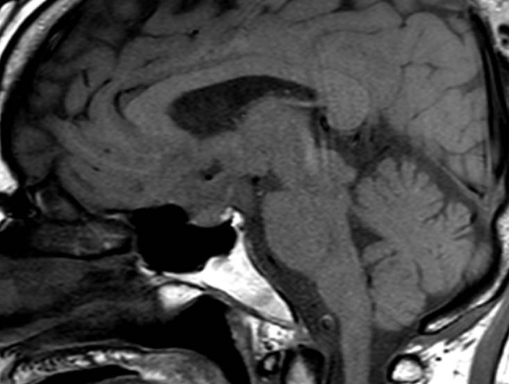
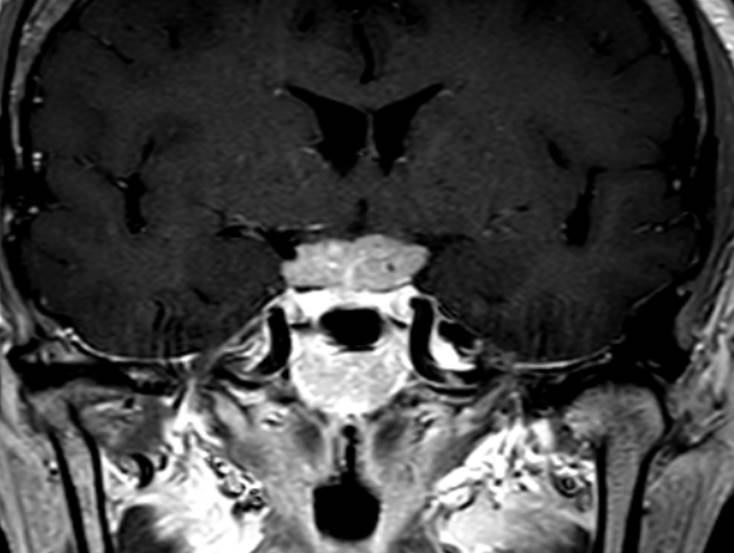
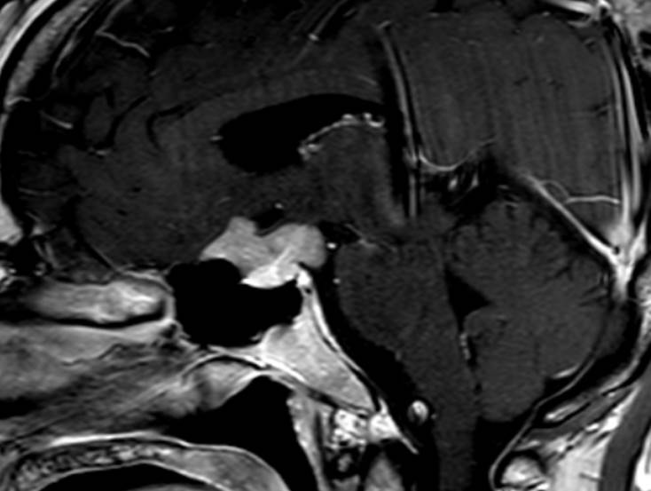
| Period | 2018-02-01~2018-02-28 |
|---|---|
| Diagnosis | Ectopic pituitary adenoma |
| Clinical information | F/56 C.C> Headache, dizziness |
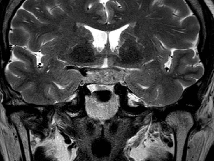
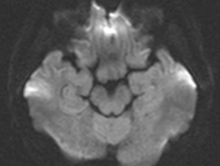
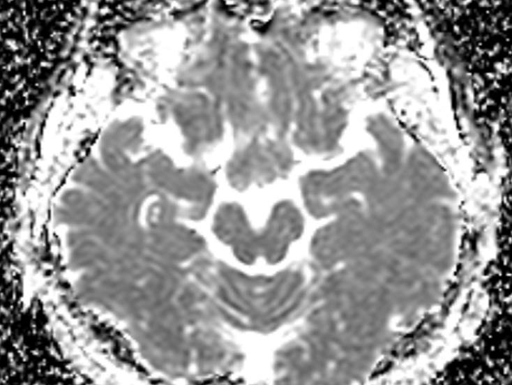
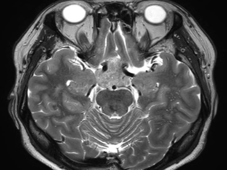
| Discussion | Ectopic pituitary adenoma MRI: -Enhancing suprasellar mass, separate from pituitary gland and pituitary stalk -T1WI: isointense signal with gray matter -T2WI: isointense to hyperintense signal intensity compared to gray matter, encasing adjacent vessels Ectopic pituitary adenoma -An adenoma of the pituitary gland located outside the sella turcica without continuity with the intrasellar normal pituitary gland -Very rare, most common in sphenoid sinus, less common in suprasellar region, cranial base, nasal cavity, cavernous sinus, 3rd ventricle, etc. -Pathologenesis: a. Adenomas derived from residual cells of Rathke's pouch to become the anterior pituitary gland, located in the sphenoid sinus or nasopharynx b. Adenomas derived from the cells of the supradiaphragmatic portion of the pars tuberalis, located in the suprasellar region c. Dissemination of the primary intrasellar adenoma (not a true ectopic pituitary adenoma) d. Adenomas derived from aberrant migrating cells of the craniopharyngeal duct in the third ventricle -Differential diagnosis: craniopharyngioma, other suprasellar lesion |
| Correct answer |
Correct Answer
Semi-Correct Answer |