HOME > Weekly Case > Weekly Case
Weekly Case
종결
Case No. 467
2018-01-01~2018-01-31
출제자 : 박지은
Hit : 259
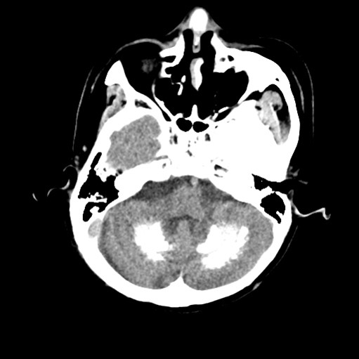
| Period | 2018-01-01~2018-01-31 |
|---|---|
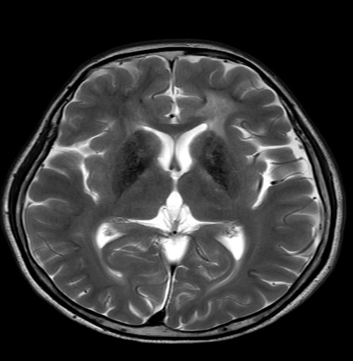
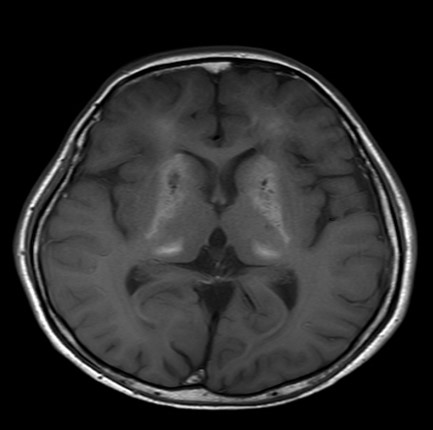
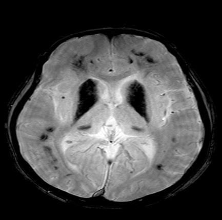
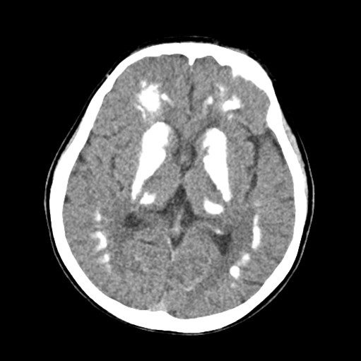
| Diagnosis | Mineralizing angiopathy |
| Clinical information | 35/M Right side weakness, dysarthria (소아때 leukemia로 치료받은 과거력) |
| Discussion | *2세에 prophylactic CNS irradiation을 받았다고 함 Parenchymal calcification within the basal ganglia and subcortical white matter Usually seen following combined radiation and chemotherapy for the treatment of CNS neoplasms in childhood Risk factor: 1) Cranial irradiation with doses as low as 1500 cGy has been associated with the development of mineralizing microangiopathy in patients with leukemia 2) The age of the patient at the time of CNS irradiation (< 10 years) appears to play a significant role Histopathology: the calcification seen in mineralizing microangiopathy is present within the walls of precapillary arterioles, capillaries, venules, and smaller arteries such as the lenticulostriate artery. Dystrophic calcification also develops in the perivascular neural tissue secondary to the mineralization of plasma fluids which leak out of the damaged vessels, and regional ischemia resulting from circulatory impairment. Reference) 1) Side effects of oncologic therapies in the pediatric central nervous system: update on neuroimaging findings https://doi.org/10.1148/rg.314105180 2) Mineralizing microangiopathy: CT and MRI. Neuroradiology (1995) 37:331-333 |
| Correct answer |
Correct Answer
Semi-Correct Answer |