HOME > Weekly Case > Weekly Case
Weekly Case
종결
Case No. 465
2017-12-01~2017-12-31
출제자 : 박미나
Hit : 272
| Period | 2017-12-01~2017-12-31 |
|---|---|
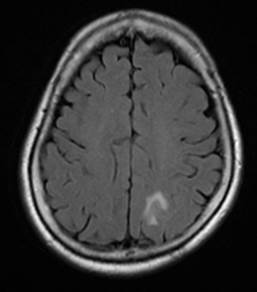
| Diagnosis | Amyloid-related imaging abnormalities associated with edema or effusion/exudate (ARIA-E) |
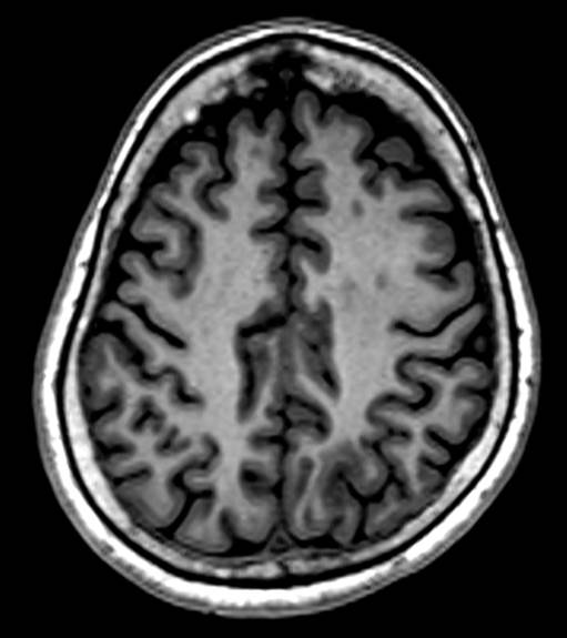
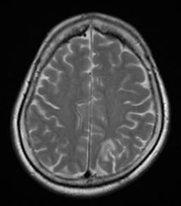
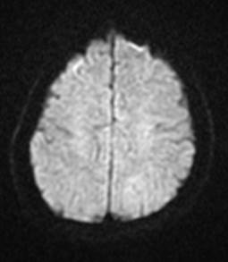
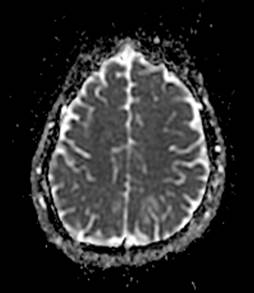
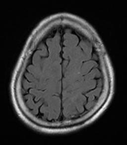
| Clinical information | F/71 Alzheimer disease 진단 후 amyloid-modifying therapy 진행 중 우연히 발견된 영상 소견 Fig 1~5는 initial Brain MRI, Fig 6는 5 months follow up MRI임. |
| Discussion | Amyloid-related imaging abnormalities associated with edema or effusion/exudate (ARIA-E) Background: Alzheimer disease (AD) remains a leading cause of morbidity and mortality without the availability of a disease-modifying agent. Several agents specifically designed to lower cerebral amyloid-beta burden have been developed and are now in various phases of clinical evaluation. Amyloid-related imaging abnormalities have been reported in the context of using amyloid-modifying therapies. Clinical Presentation: It has typically been identified in asymptomatic patients undergoing routine scheduled MR imaging surveillance Key Diagnostic Feature: ARIA-E: FLAIR signal abnormalities thought to represent parenchymal edema and/or sulcal effusions (ie, exudates) ARIA-H: hemosiderin deposition detected on T2*-weighted sequences believed to represent blood-degradation products, including microhemorrhages (10mm) and larger superficial hemosiderin deposits (10 mm). |
| Correct answer |
Correct Answer
Semi-Correct Answer |