HOME > Weekly Case > Weekly Case
Weekly Case
종결
Case No. 452
2017-08-01~2017-08-31
출제자 : 강경미
Hit : 310
| Period | 2017-08-01~2017-08-31 |
|---|---|
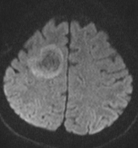
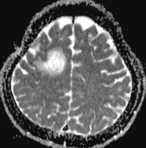
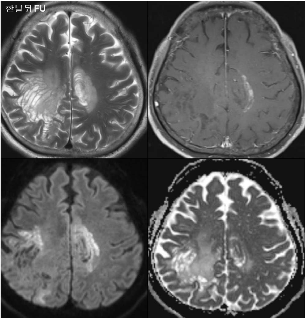
| Diagnosis | Balo’s concentric sclerosis |
| Clinical information | M/32 Left motor weakness (1 month ago) 진단은? |
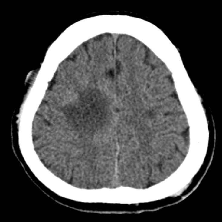
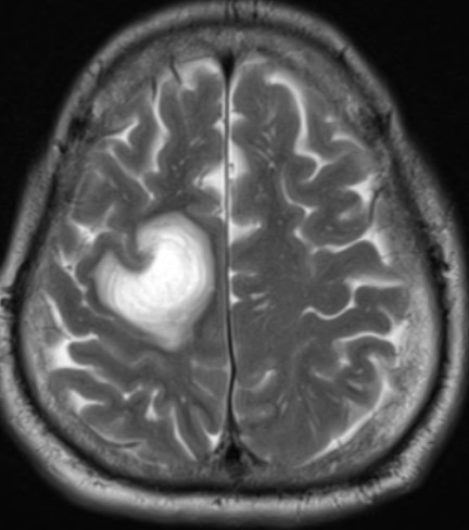
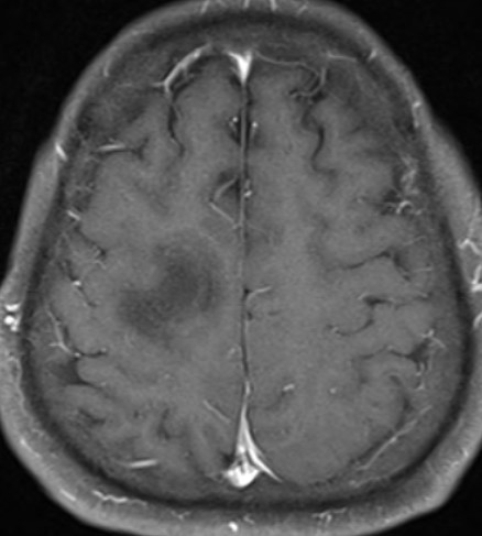
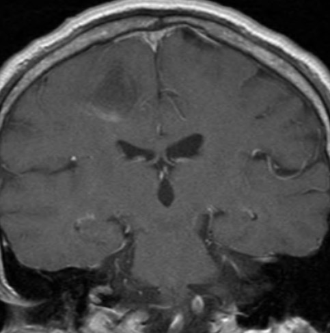
| Discussion | Balo’s concentric sclerosis • Rare demyelinating disease, considered as a variant of multiple sclerosis • Clinical presentation – Acute or subacute neurological deterioration, sometimes fulminant course – Can be mistaken for a brain tumor – MR imaging features – T1 : Alternating iso- and hypointense concentric rings – T2 : Hyperintense concentric lamellae – Location : Predominanty cerebral WM – BG, pons, cerebellum, spinal cord and optic nerve involvement has been reported – DWI : High SI at the lesion edge, without definite low ADC value – Gd Enhancement : peripheral enhancement / multiple layers corresponding to T2 high layers • FDG-PET uptake (-) DDx from other acute demyelinating ds or high-grade tumors • CSF study : Oligoclonal bands may be present • Pathology – Consists of cerebral WM oligodendrocyte loss and demyelination (similar to MS) – Sparing of the cortical gray matter (unlike conventional MS) – Alternating ring appearance : Alternating areas of relative myelin preservation and loss Areas of relative myelin preservation : Early or partial demyelination(not normal myelin) • Immunopathogenesis for concentric lesions • Arise around a perivenular zone in response to an unknown stimulus Macrophages and activated microglia : Cytokine, oxygen free radicals, other mediators induce demyelination Hypoxia-ischemia may also contribute to damage – Concentric rings : Damage from the successive outward spread of chemical mediators in waves from a focal point Reference> Lancet Neurol 2014; 13: 740–46 |
| Correct answer |
Correct Answer
Semi-Correct Answer |