HOME > Weekly Case > Weekly Case
Weekly Case
종결
Case No. 451
2017-07-01~2017-07-31
출제자 : 이호준
Hit : 322
| Period | 2017-07-01~2017-07-31 |
|---|---|
| Diagnosis | X-linked adrenoleucodystrophy (ALD), anterior white matter pattern |
| Clinical information | M/11 Past History: IUP 40wks, 3.2kg, NSVD, no peri/post natal history 진단명? |
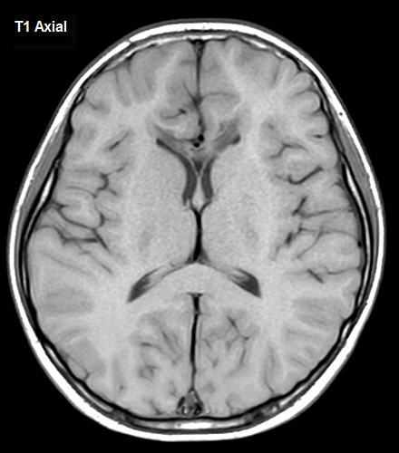
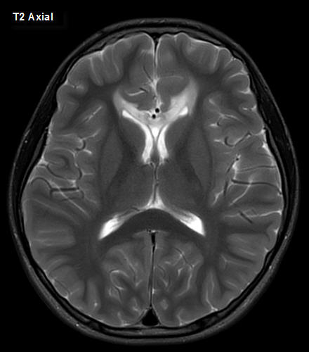
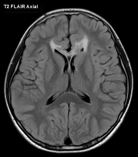
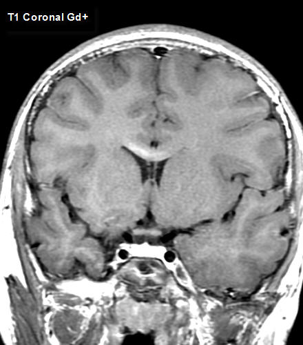
| Discussion | X-linked adrenoleucodystrophy (ALD), anterior white matter pattern Family history: Uncle X-linked ALD, mother and grandmother carrier of ABCD1 mutation Lab: Increased ACTH levels (421.20 pg/mL; normal range 7.2~63.3 pg/mL) On T1, T2, weighted and T2 weighted FLAIR images, increased T2 signal, and decreased T1 signal is seen in the genu of corpus callosum, and adjacent frontal white matter. Contrast enhancement is seen at the anterior body of corpus callosum, posterior to the previous described findings. The patient was screened for ABCD1 gene mutation, which was positive. = Bronze Shilder disease -X-linked recessive inherited disorder of peroxisome metabolism : impaired β-oxidation of very long chain fatty acids with accumulation in white matter / Mutations of ABCD1 gene -Phenotypic variability even in same family -Innermost zone consists of necrosis gliosis with or without calcification, intermediate zone of active demyelination and inflammation, peripheral zone of demyelination without inflammation * Imaging findings A. MRI/CT Decreased T1 signal, Increased T2 signal of involved WM Typical childhood cerebral X-ALD : splenium, peritrigonal WM, corticospinal tract, fornix, commissural fibers, visual and auditory pathways (asymmetric progression toward frontal or temporal lobe, central to peripheral) Usually bilateral but rarely unilateral Anterior white matter pattern in up to 15% Frontal white matter, genu of corpus callosum, frontopontine tract …… Enhancement of intermediate zones (leading edge) Contrast enhancement correlates with likelihood of progression Restricted diffusion in involved white matter Calcification may be present References 1. Diagnostic Imaging, Brain, Second Edition, 2010, AMIRSYS |
| Correct answer |
Correct Answer
Semi-Correct Answer |