HOME > Weekly Case > Weekly Case
Weekly Case
종결
Case No. 450
2017-07-01~2017-07-31
출제자 : 이호준
Hit : 312
| Period | 2017-07-01~2017-07-31 |
|---|---|
| Diagnosis | Lymphocytic hypophysitis |
| Clinical information | F/45 CC: Amenorrhea (x 2 years), visual disturbance Lab: Hypopituitarism |
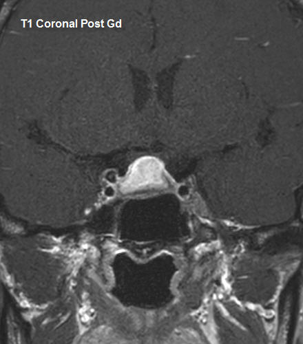
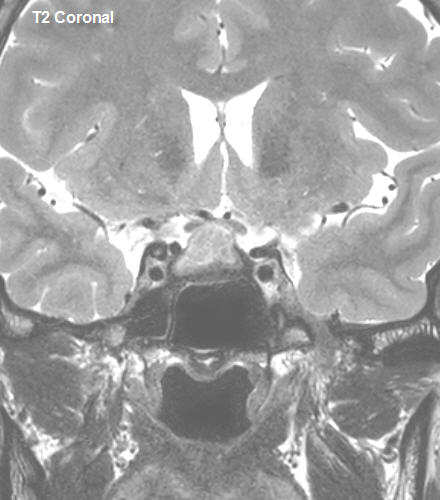
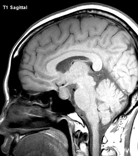
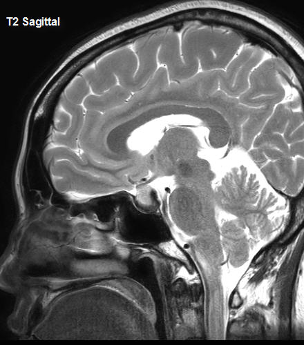
| Discussion | Pituitary height/volume is increased with suprasellar extension and optic chiasm bowing. On coronal and sagittal T2 weighted image, a dark intensity rim is seen around the pituitary gland. The corresponding area shows poor enhancement on coronal T1 post Gd enhancement. The pituitary gland shows strong homogenous enhancement. On sagittal T1 weighted image, the normal T1 hypersignal intensity of the neurohypophysis lost, but no symptoms of diabetes insipidus was present. Stalk assessment was difficult due to compression, but thickening was not definite. On surgery, a solid fibrotic mass with adhesion was noted, and pathologic findings of multifocal lymphoid follicle formation were reported. = Adenohypophysitis, primary hypophysitis, stalkitis -Rare disease (1-2% of sellar lesions) -Mean age F=35 years, M=45 years, M:F = 1:8-9. -Most common presentation is headache or visual impairment -Hypopituitarism may be frequently be present -Clinical and radiologic features may mimic adenoma. Differentiation is important since treatment options are different. * Imaging findings A. MRI Enlarged pituitary gland Uniform, intense contrast enhancement Loss of posterior pituitary T1 hyperintensity Pituitary Symmetry Stalk thickening “Parasellar T2 Dark Sign” Characteristic sign : Low sensitivity, high specificity May reflect fibrotic changes with infiltration of lymphoplasmacytic cells * Differential Diagnosis - Pituitary hyperplasia, pituitary adenoma, metastasis, lymphoma, sarcoidosis, References 1. Diagnostic Imaging, Brain, Second Edition, 2010, AMIRSYS 2. Nakata, Y., et al. "Parasellar T2 dark sign on MR imaging in patients with lymphocytic hypophysitis." American Journal of Neuroradiology 31.10 (2010): 1944-1950. |
| Correct answer |
Correct Answer
Semi-Correct Answer |