HOME > Weekly Case > Weekly Case
Weekly Case
종결
Case No. 449
2017-07-01~2017-07-31
출제자 : 이호준
Hit : 272
| Period | 2017-07-01~2017-07-31 |
|---|---|
| Diagnosis | Acute Calcific tendinitis(tendonitis) of longus colli |
| Clinical information | M/39 CC: Odynophagia, neck pain x 2 weeks Lab: WBC 12610, ESR/CRP 29/68.2, BT 37.3 |
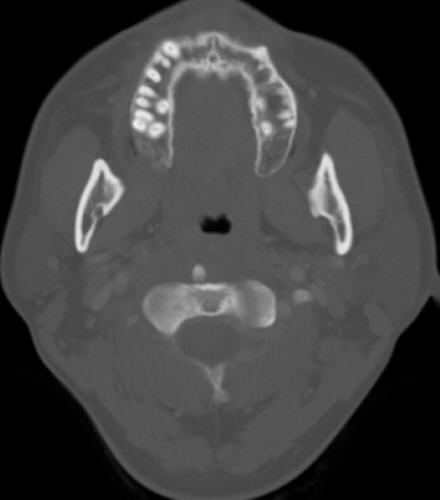
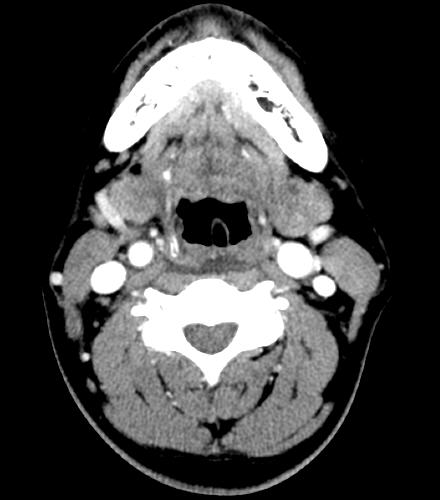
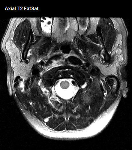
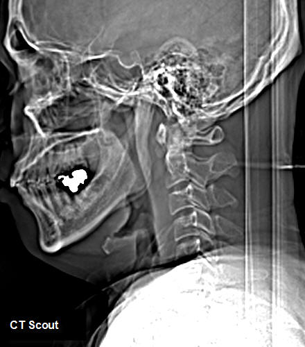
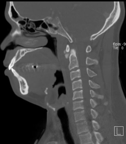
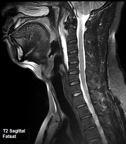
| Discussion | 해설: On CT scout image, widened pre-vertebral soft tissue is seen with calcification anterior to C2. Fat saturated T2 axial and sagittal scan shows increased signal in the longus colli muscle, with retropharyngeal edema and fluid collection. Low signal is seen anterior to the dens on MRI, which is shown to be calcifications, on bone setting of sagittal reformatted and axial neck CT images. Contrast enhanced CT scan shows retropharyngeal fluid collection, without definite contrast enhancement. = retropharyngeal calcific tendinitis, acute calcific pre-vertebral tendonitis -Under-recognized cause of spontaneous acute or subacute neck pain, dysphagia, or odynophagia -Affecting patients during 3rd ~ 6th decades of life -Mild elevation of ESR/CRP and WBC count, low grade fever may be present -Deposition of calcium hydroxyapatite crystals induce a local acute inflammatory response fluid collection within the retropharyngeal space -Recognition of this uncommon presentation is important to prevent unnecessary surgical incision and drainage since majority of this disease is managed conservatively, with NSAIDs and rest. * Imaging findings A. MRI/CT Focal C1-2 pre-vertebral calcification (variable degree, may be subtle; pathognomic) Smooth, expanding retropharyngeal space edema without rim enhancement Signal change of longus colli on MRI B. X-ray Widening of pre-vertebral soft tissue, occasional visualization of calcification * Differential Diagnosis - Retropharyngeal space effusion not associated with longus colli tendinitis, retropharyngeal abscess, pre-vertebral space infection References 1. Diagnostic Imaging, Head and Neck, Second Edition, 2011, AMIRSYS 2. Offiah, C. E., and E. Hall. "Acute calcific tendinitis of the longus colli muscle: spectrum of CT appearances and anatomical correlation." The British journal of radiology 82.978 (2009): e117-e121. |
| Correct answer |
Correct Answer
Semi-Correct Answer |