HOME > Weekly Case > Weekly Case
Weekly Case
종결
Case No. 443
2017-05-01~2017-05-31
출제자 : 이아름
Hit : 346

| Period | 2017-05-01~2017-05-31 |
|---|---|
| Diagnosis | Wernicke encephalopathy |
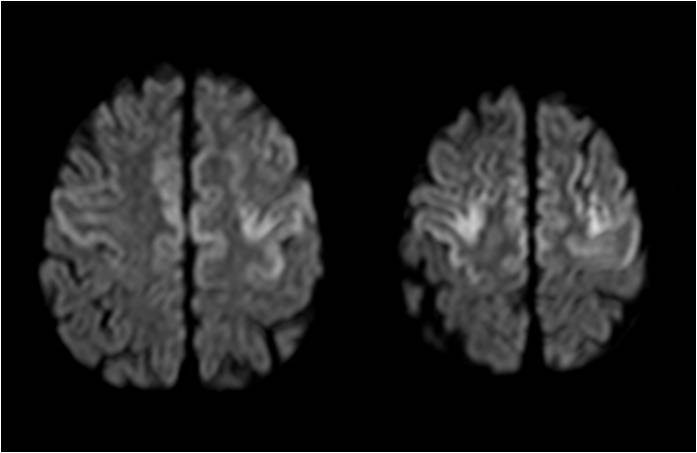
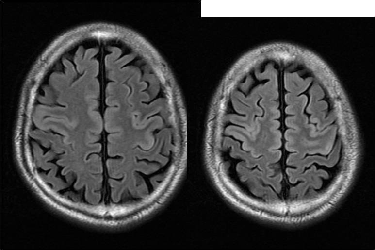
| Clinical information | 53/M C.C: mental change Clinical history: HTN, DM, Chronic alcoholism, Poor nutrition Fig 1A, 1B: initial MRI (the day of admission) Fig 1C: FU MRI after 1 day |
| Discussion | The patient on quiz was treated with Thiamine supplement. After 1 day, FU MRI showed decrease in extent of T2 high SI lesion involving bilateral cerebral cortex (mainly in pre-central sulcus) Wernicke encephalopathy : Typical MRI findings include the symmetrical involvement of the bilateral medial thalami, mammillary bodies and periaqueductal gray matter Atypical manifestation including involvement of the cerebellum, (including the vermis), cranial nerve nuclei, red nuclei, dentate nuclei, splenium of corpus callosum and cerebral cortex Reference journal: A Case of Atypical Wernicke’s Encephalopathy Showing Symmetric Cortical Lesions on MRI, J Neurocrit Care 2013;6:43-46 |
| Correct answer |
Correct Answer
Semi-Correct Answer |