HOME > Weekly Case > Weekly Case
Weekly Case
종결
Case No. 433
2017-01-01~2017-01-31
출제자 : 박지은
Hit : 418
| Period | 2017-01-01~2017-01-31 |
|---|---|
| Diagnosis | Nodular fasciitis |
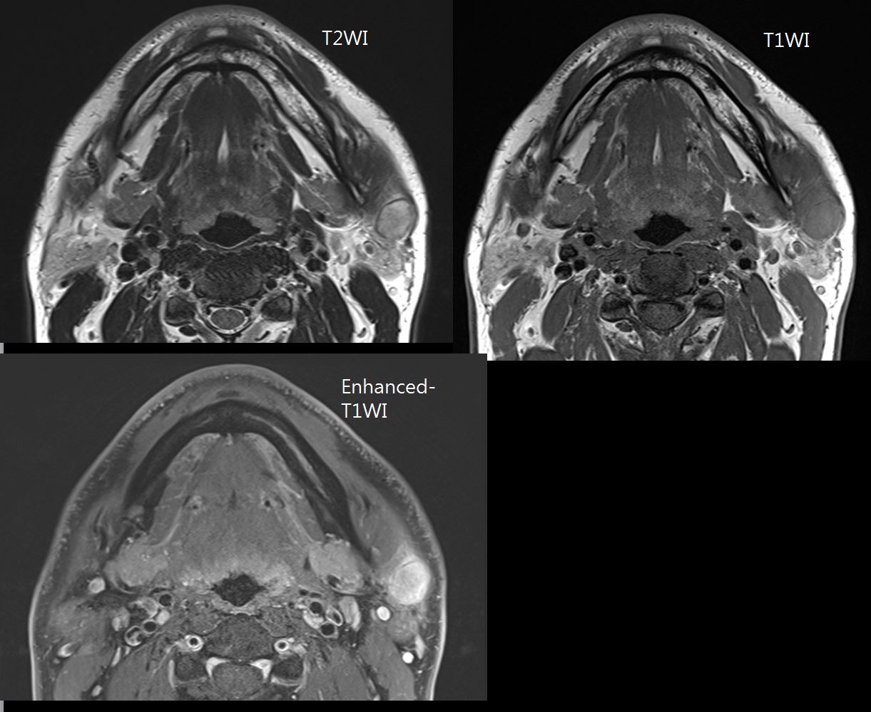
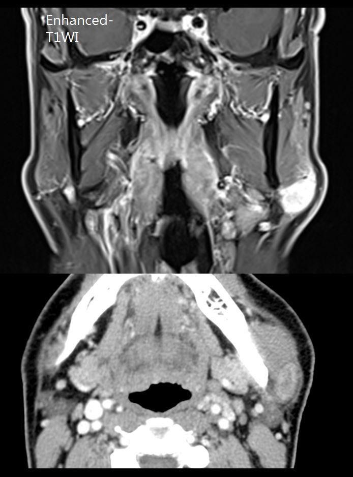
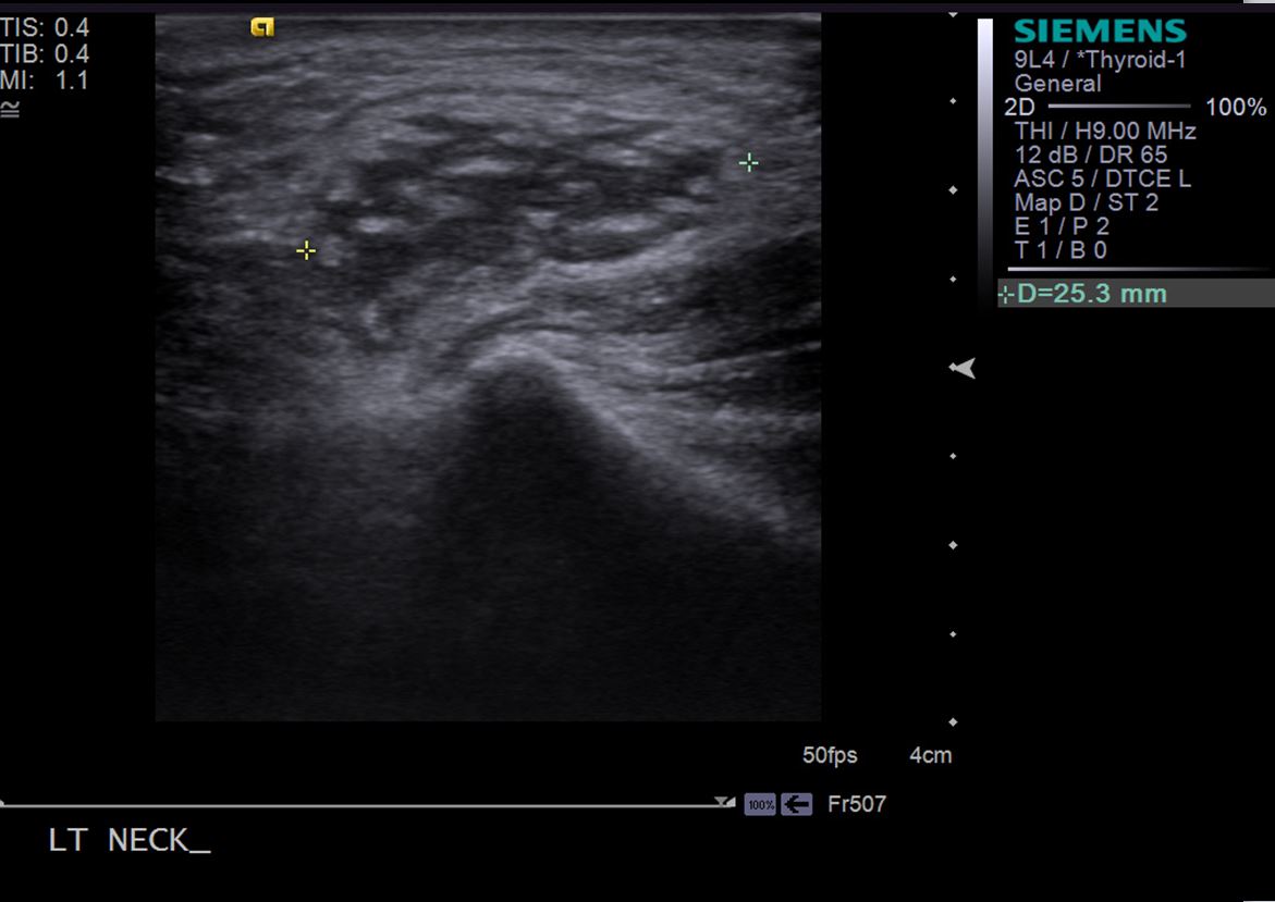
| Clinical information | Left palpable neck mass를 주소로 내원한 55세 남자환자이다. 진단은? |
| Discussion | Nodular fasciitis 해설: • a benign proliferation of fibroblasts and myofibroblasts that may be mistaken for a sarcomatous lesion • most often occurs in patients between 20 and 40 years of age • subcutaneous, fascia based, and circumscribed • types: myxoid, cellular, or fibrous, according to the predominant histologic feature of the lesion • Radiological features – Contrast enhancement is typically diffuse but may be peripheral – hypercellular lesions appears nearly isointense to that in skeletal muscle on T1-weighted images – hyperintense to that in adipose tissue on T2- weighted images – Reference) Haldorsen IS, Espeland A, Larsson EM. AJNR 2011;32 (6): 984-92. DDx) Kimura disease - Asian males who are in their 2nd or 3rd decade of life. - head and neck and often involve the major salivary glands - bulky, poorly marginated subcutaneous soft-tissue masses with signal that is isointense to or slightly higher in intensity than the signal of muscle on T1-weighted images - The masses are composed of enlarged lymph nodes with eosinophilic infiltration in the paracortical areas, increased postcapillary venules, and hyperplastic lymphoid follicles |
| Correct answer |
Correct Answer
Semi-Correct Answer |