HOME > Weekly Case > Weekly Case
Weekly Case
종결
Case No. 431
2017-01-01~2017-01-31
출제자 : 박지은
Hit : 396

| Period | 2017-01-01~2017-01-31 |
|---|---|
| Diagnosis | Cortical dysplasia |
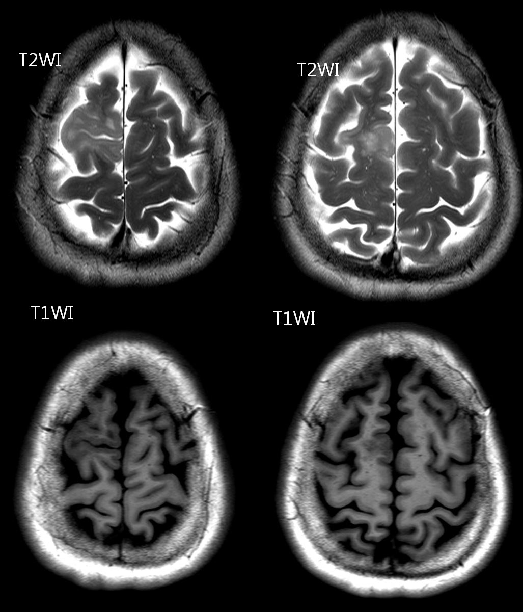
| Clinical information | Dizziness 를 주소로 내원한 53세 여자환자이다. 진단은? |
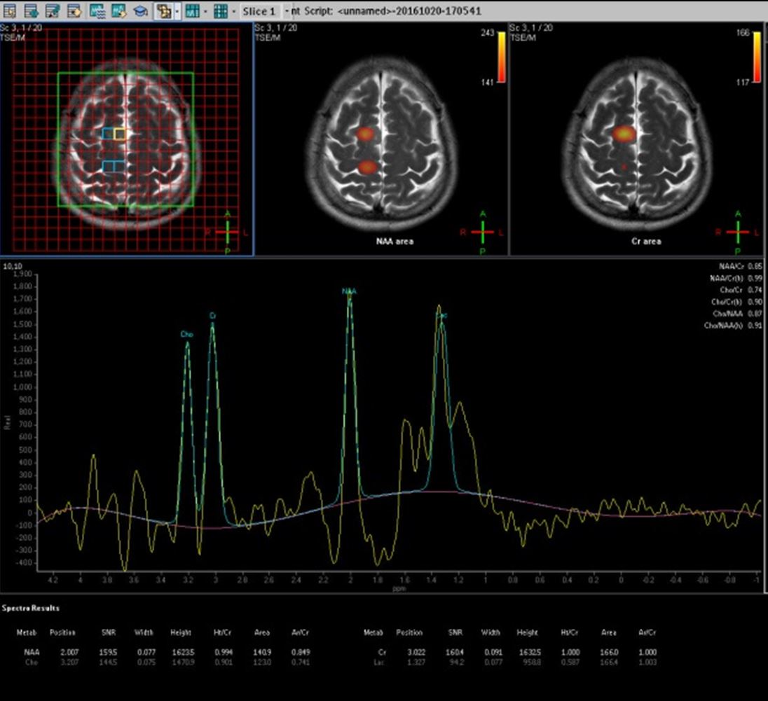
| Discussion | Cortical Dysplasia 해설: 본 증례는 병원 내에서 low grade glioma와 함께 감별이 되었던 증례입니다. Cortical thickening은 있으나 white matter volume이 감소되어 있고, superior frontal sulcus가 left side와 asymmetric 한 점, MR spectroscopy 소견이 감별에 도움이 되겠습니다 Heterogenous groups of disorders or cortical formation Architectural and proliferative features MRI) -Cortical thickening -Blurring of white matter-grey matter junction with abnormal architecture of subcortical layer -T2/FLAIR hyperintensity of white matter, grey atter -Abnormal sulcal or gyral pattern Blumke classification (2011) 1) Type I : Ia usually confined to temporal lobes Ib outside of temporal lobes Blurring of grey/white matter junction, segmental lobar atrophy with loss of regional white matter volume 2) Type II : Frontal lobes Blurring of grey/white matter junction, cortical thickening, transmantle sign 3) Type III : adjacent other abnormalities (IIIa-hippocampal atrophy, IIIb-glioneural tumor, IIIc-vascular malformation, IIId-early childhood insult(gliosis) Reference) Barkovich AJ, Guerrini R, Kuzniecky RI et al Brain. 2012;135 (5): 1348-69. Ddx > Low grade glioma |
| Correct answer |
Correct Answer
Semi-Correct Answer |