HOME > Weekly Case > Weekly Case
Weekly Case
종결
Case No. 430
2016-12-01~2016-12-31
출제자 : 배윤정
Hit : 445
| Period | 2016-12-01~2016-12-31 |
|---|---|
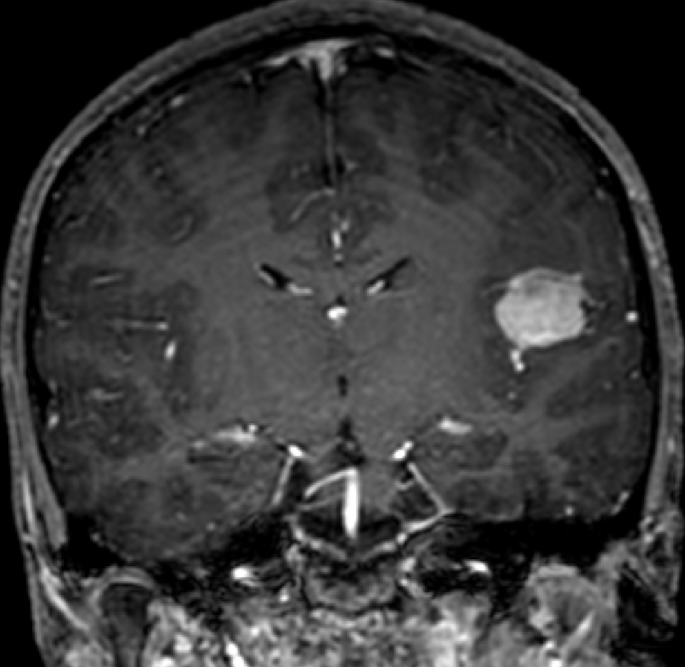
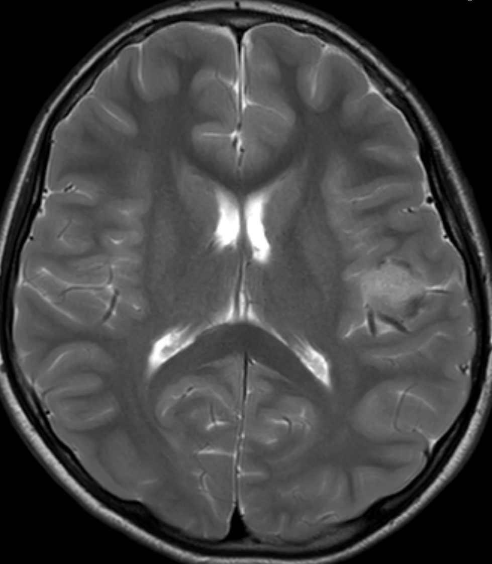
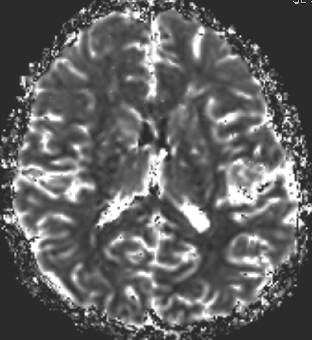
| Diagnosis | Meningioma, intraparenchymal |
| Clinical information | M/13 PI> Headache |
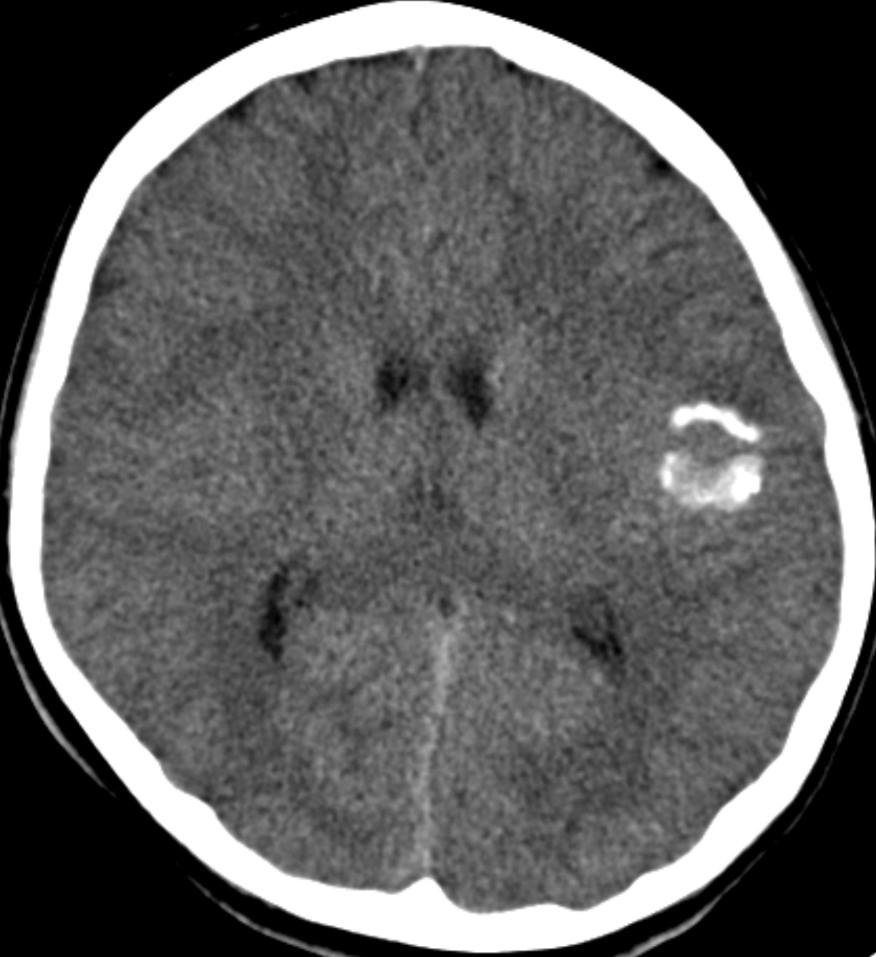
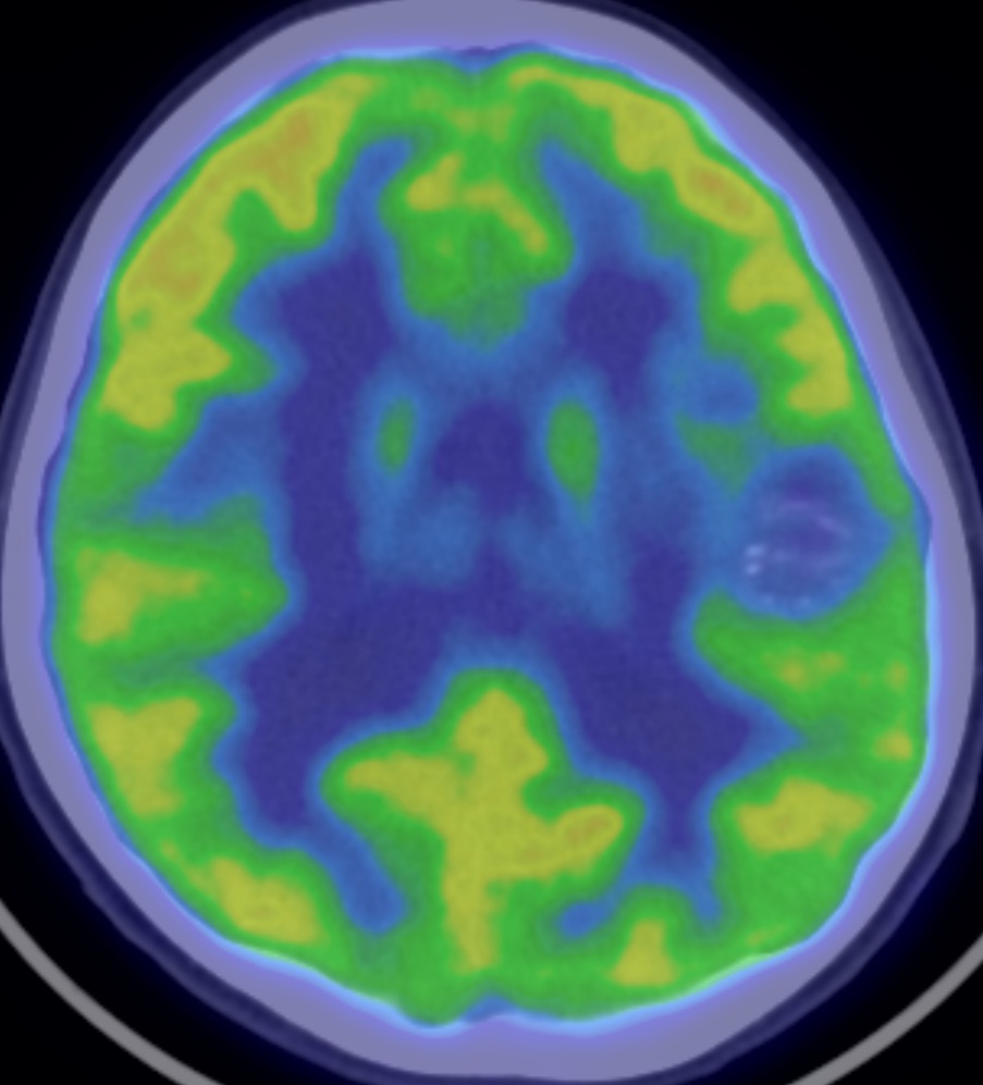
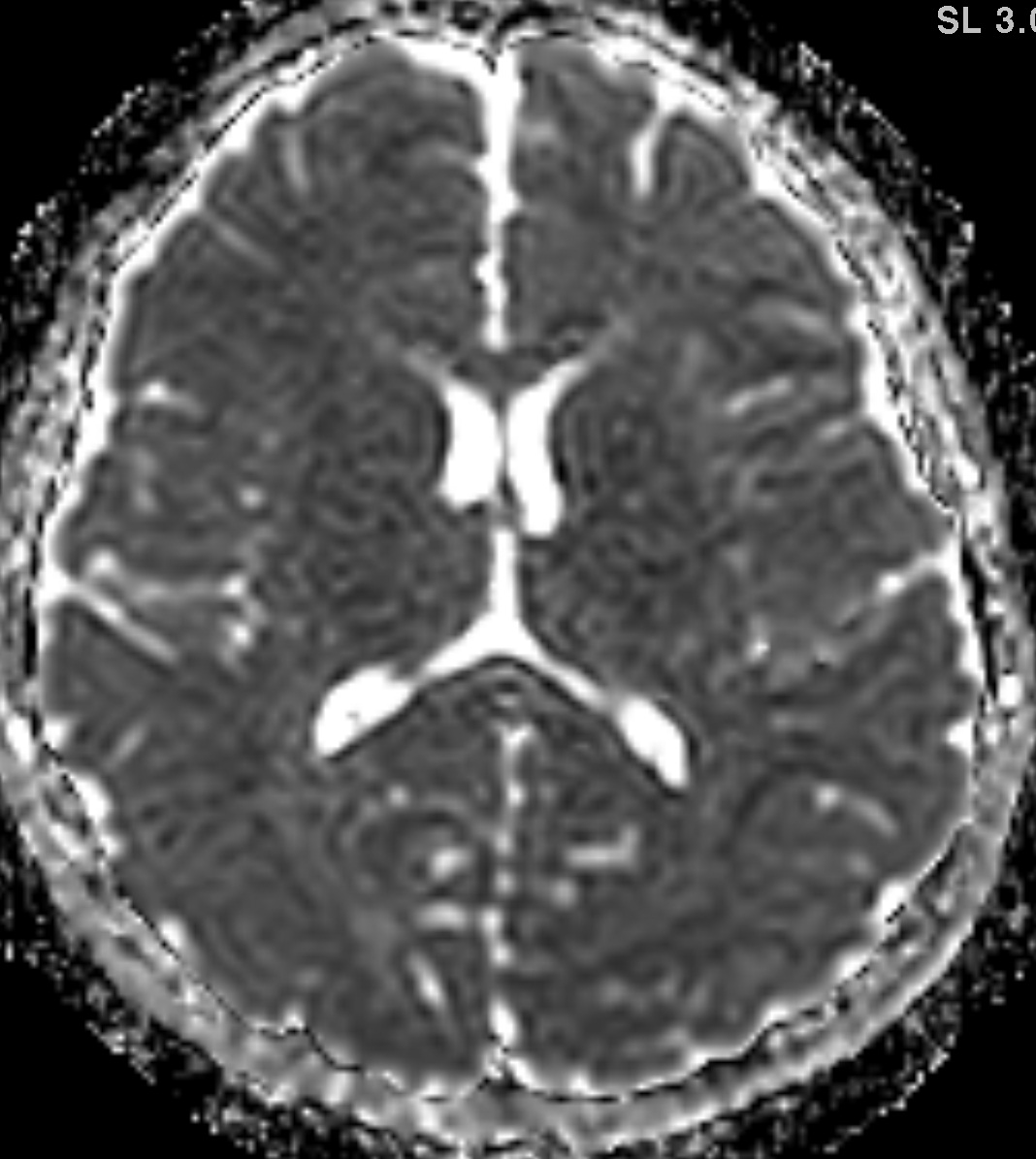
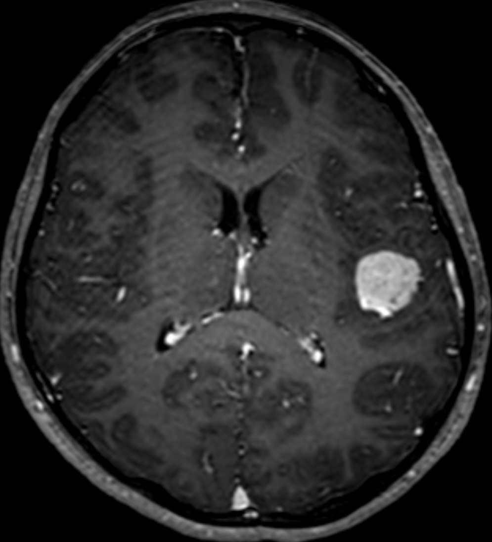
| Discussion | Meningioma, intraparenchymal CT: About 2cm sized mass with calcification MRI: About 2cm sized mass with avid enhancement, no dural attachment, ADC similar to gray matter, mildly increased CBV PET: Hypometabolism on FDG PET Pathology: Intraparenchymal meningioma with 1) diffuse expression of Vimentin 2) equivocal expression of S100 protein 3) no expression of GFAP, EMA and Synaptophysin (no evidence of glioma and neuronal tumors) 4) Ki-67 proliferative index : 4% Intraparenchymal meningioma -Intraparenchymal meningiomas are believed to arise from arachnoid cells of the pia matter, which supplies the perforating blood vessels as they enter the surface of brain or sulci. -The key feature in the neuroimaging-based diagnosis of intraparenchymal compared with dural meningioma is the absence of dural attachment. -Pediatric meningioma a. About 2% of meningiomas occur in children, especially in the 10–15 y/o age group (62%) b. Meningiomas without dural attachment: intraventricular, pineal region, deep sylvian, intraparenchymal/subcortical, etc. c. Sylvian/intraparenchymal tumors often have feeding arteries from MCA. |
| Correct answer |
Correct AnswerSemi-Correct Answer |