HOME > Weekly Case > Weekly Case
Weekly Case
종결
Case No. 429
2016-12-01~2016-12-31
출제자 : 배윤정
Hit : 375
| Period | 2016-12-01~2016-12-31 |
|---|---|
| Diagnosis | Amyloidosis |
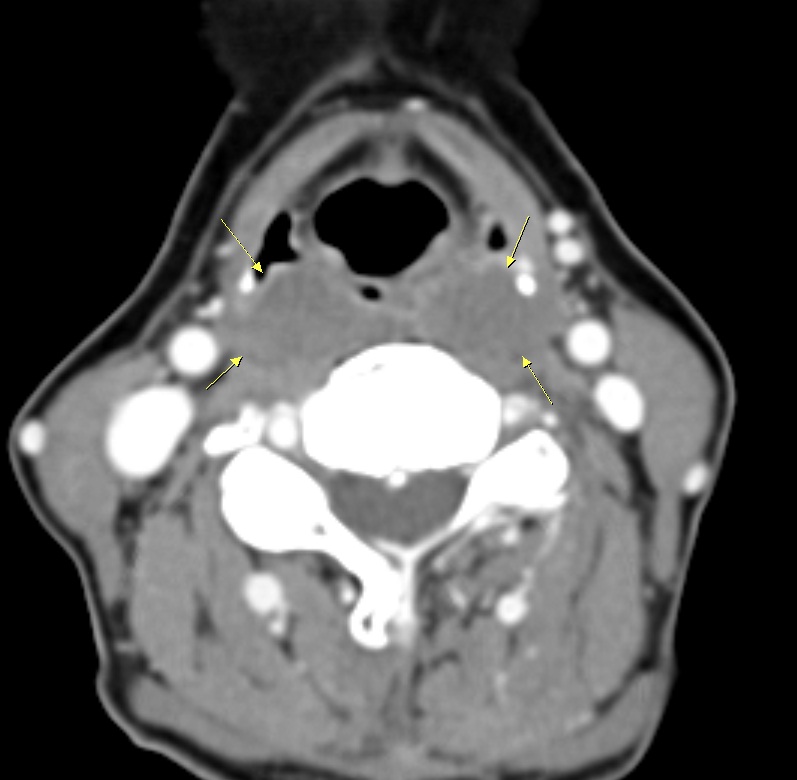
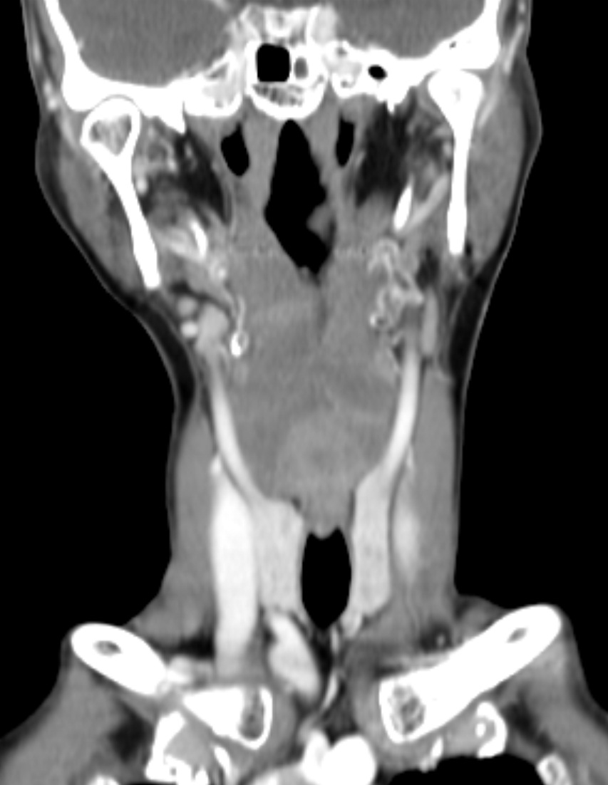
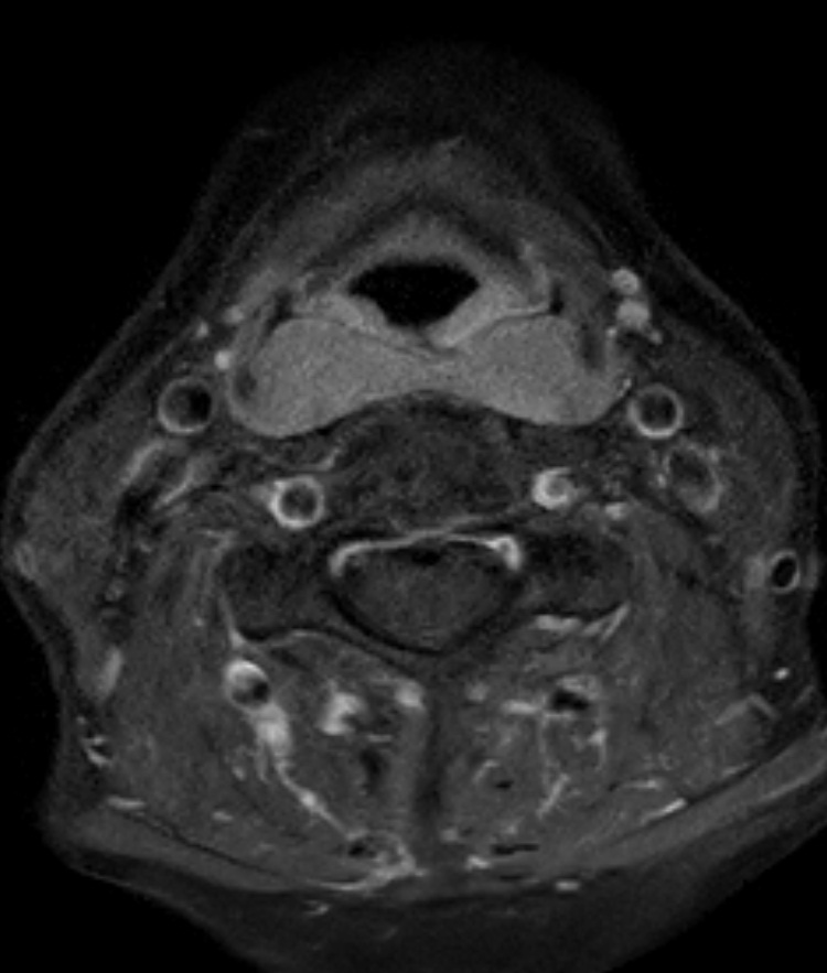
| Clinical information | F/73 PI>Dysphagia, throat discomfort Laryngoscopy: pharyngeal wall bulging and swelling |
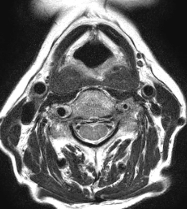
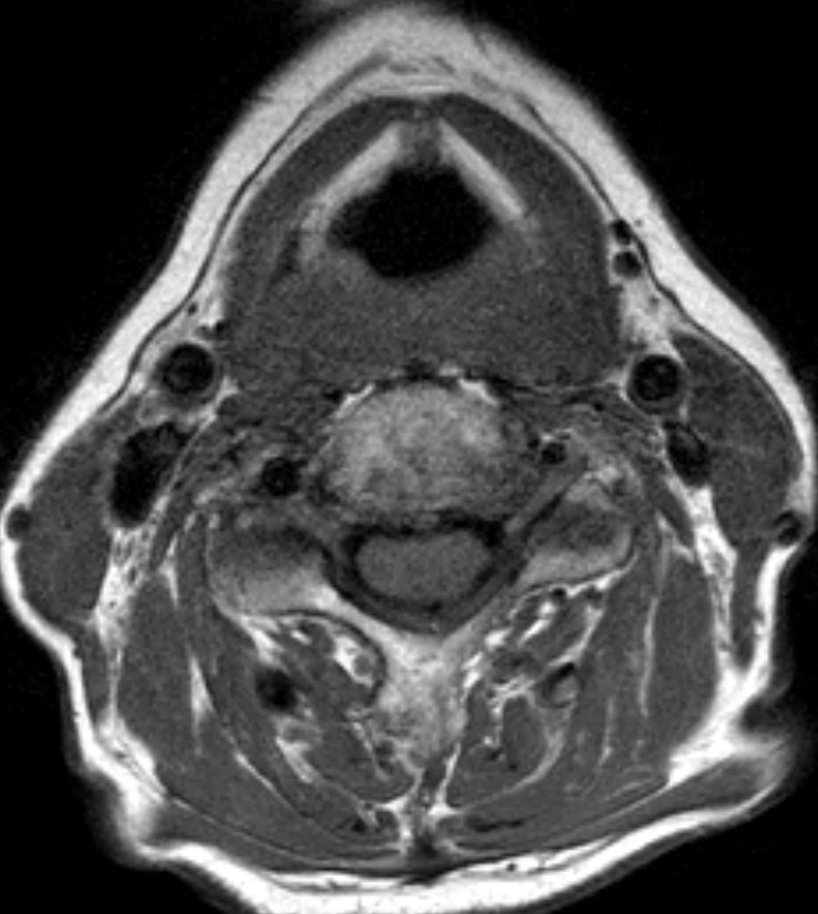
| Discussion | Amyloidosis CT: Diffuse submucosal soft-tissue lesion around the posterior pharynx. MRI: Diffuse submucosal soft-tissue lesion around the posterior harynx, showing typical low T2-weighted signal intensity, and low T1-weighted signal intensity, paralleling the signal intensities of skeletal muscle Amyloidosis -Amyloidosis is characterized by infiltration and extracellular deposition of homogeneous, eosinophilic proteinaceous fibrils in a β-pleated sheet configuration. It classically has an apple-green birefringence when stained with Congo red and viewed under polarized light. -Classification a. Localized disease: involves only one organ, typically the larynx or brain (where it has been associated with aging and Alzheimer’s disease) b. Systemic amyloidosis b-1. Primary amyloidosis: spontaneous overproduction of the antibody light chain in the bone marrow plasma cell b-2. Secondary amyloidosis: result of an underlying condition such as multiple myeloma, chronic infections (tuberculosis or osteomyelitis), and chronic inflammatory diseases such as rheumatoid arthritis, Sjögren’s disease, and ankylosing spondylitis b-3. Familial type: autosomal dominant pattern of inheritance with amyloid deposits composed of the protein transthyretin, which is made in the liver -Most common head and neck sites: supraglottic and subglottic larynx, thyroid gland, and rarely pharynx (nasopharynx more often than oropharynx or hypopharynx) -Imaging a. MR imaging is the modality of choice b. Typically, the amyloid deposits have an intermediate T1-weighted signal intensity and a low T2-weighted signal intensity, similar to skeletal muscle. This is secondary to the β-pleated structure of the amyloid deposits that occur as protein fibrils in a parallel sheet-like configuration, similar to the organization of skeletal muscle fibers. |
| Correct answer |
Correct AnswerSemi-Correct Answer |