HOME > Weekly Case > Weekly Case
Weekly Case
종결
Case No. 425
2016-11-01~2016-11-30
출제자 : 유인선
Hit : 426
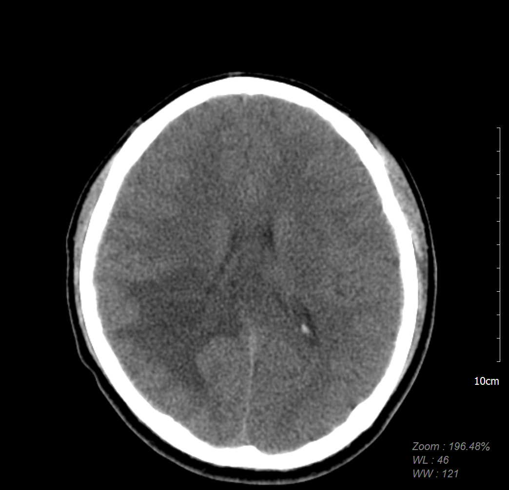
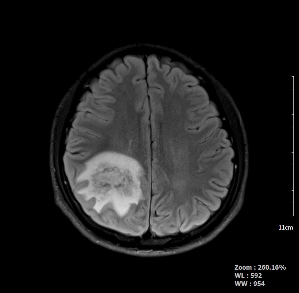

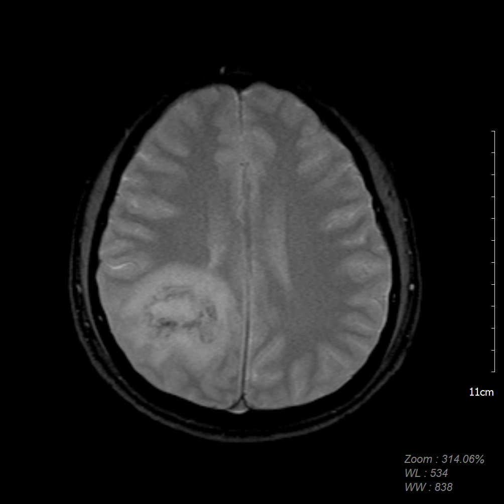
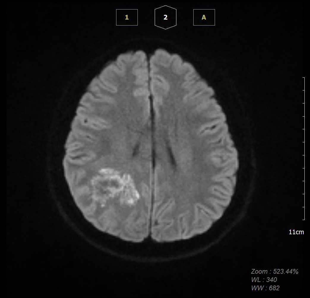
| Period | 2016-11-01~2016-11-30 |
|---|---|
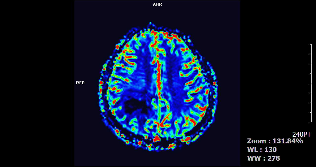
| Diagnosis | CNS vasculitis (Lymphohistiocytic venulitis with occasional fibrin thrombi) |
| Clinical information | M/20 C.C> 5일전부터 시작된 headache |
| Discussion | CNS vasculitis: necrosis of blood vessel walls. Both arteries, veins may be affected Imaging findings MR: BG, cortex, subcortical WM: multifocal hypodensities, T2 hyperintensities with or without secondary findings (ischemia, infarction), may show hemorrhages on GRE, can show restricted diffusion on DWI in acute phase, and may see patchy enhancement Best Imaging tool: HR contrast enhanced MR with vessel wall imaging |
| Correct answer |
Correct AnswerSemi-Correct Answer |