HOME > Weekly Case > Weekly Case
Weekly Case
종결
Case No. 421
2016-09-01~2016-09-30
출제자 : 정우상
Hit : 420
| Period | 2016-09-01~2016-09-30 |
|---|---|
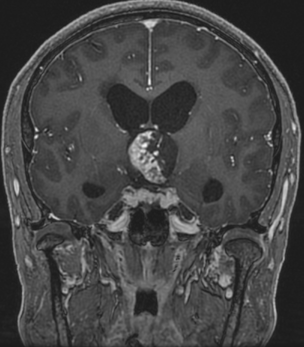
| Diagnosis | Adamantinomatous craniopharyngioma in the 3rd ventricle |
| Clinical information | M/51 C.C> Blurred vision x 5months 진단은? |
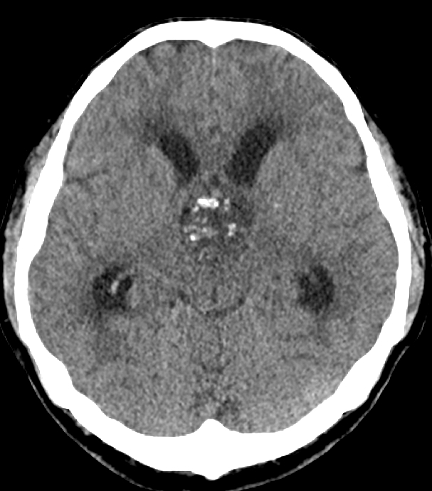
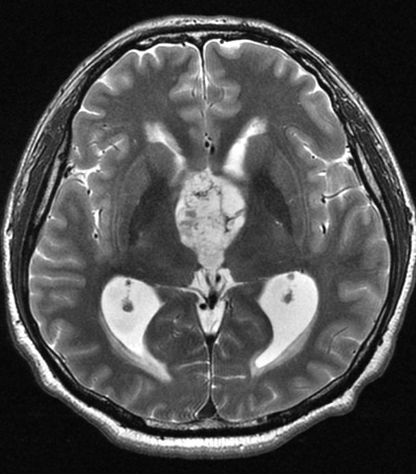
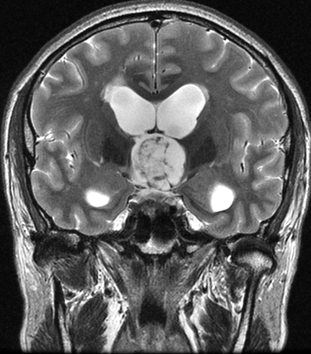
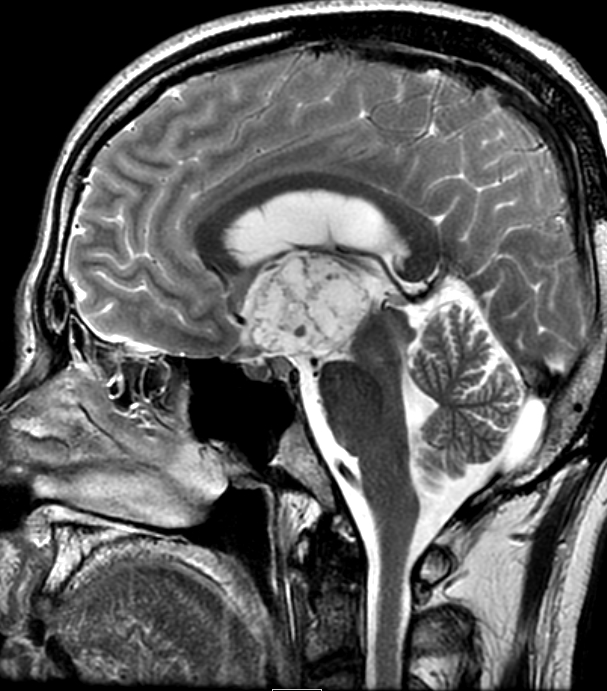
| Discussion | About 4x3cm bubbly appearance solid and cystic mass in 3rd ventricle. Calcification on CT scan. <해설> In the vast majority of cases, craniopharyngiomas have a large suprasellar component (95%), with most involving both the suprasellar and intrasellar spaces (75%). A minority are purely suprasellar (20%), whereas purely intrasellar location is quite uncommon (<5%), and may be associated with the expansion of the pituitary fossa. Occasionally, craniopharyngiomas appear as intraventricular, homogeneous, soft-tissue masses without calcification (papillary subtype). The third ventricle is a particularly common location. Rare or ectopic locations reported include: nasopharynx, posterior fossa, extension down the cervical spine. |
| Correct answer |
Correct Answer
Semi-Correct Answer |