HOME > Weekly Case > Weekly Case
Weekly Case
종결
Case No. 416
2016-08-01~2016-08-31
출제자 : 강경미
Hit : 358
| Period | 2016-08-01~2016-08-31 |
|---|---|
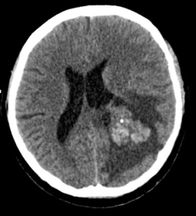
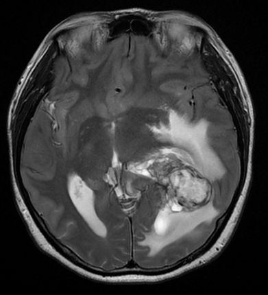
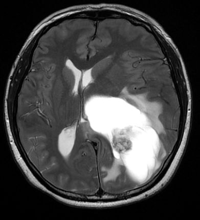
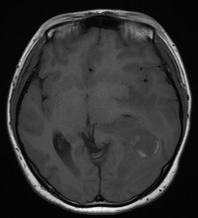
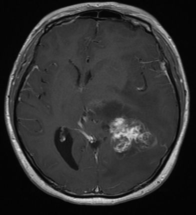
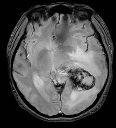
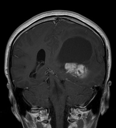
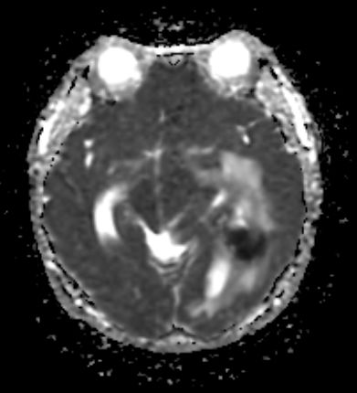
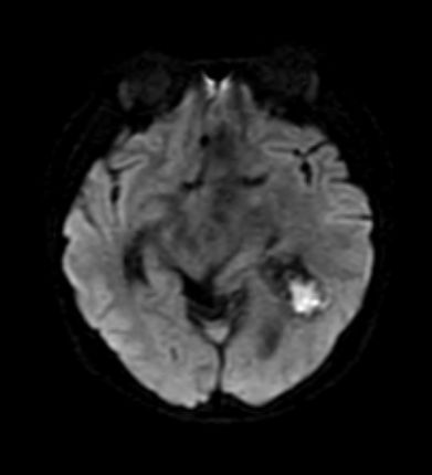
| Diagnosis | Cavernous malformation |
| Clinical information | F/30 두통 진단은? |
| Discussion | Benign vascular hamartoma Contains masses of closely apposed immature blood vessels ("caverns"), no neural tissue Intralesional hemorrhages of different ages CMs exhibit range of dynamic behaviors (enlargement, regression, de novo formation) Imaging CMs vary from microscopic to giant (> 6 cm) Locules of variable size contain blood products at different stages of evolution Variable appearance depending on hemorrhage/stage CT If large, a region of hyperdensity can be seen. If there has been a recent bleed, it is more conspicuous and may be surrounded by a mantle of edema. MRI “popcorn” or “berry” appearance with a rim of signal loss due to hemosiderin Prominent blooming on SWI T1 and T2 WI : vary (depending on the age of the blood produces) Reference> Diagnostic Imaging: Brain, Osborn et al, Amirsys, 2009 |
| Correct answer |
Correct Answer
Semi-Correct Answer |