HOME > Weekly Case > Weekly Case
Weekly Case
종결
Case No. 415
2016-07-01~2016-07-31
출제자 : 안성수
Hit : 403
| Period | 2016-07-01~2016-07-31 |
|---|---|
| Diagnosis | Colloid cyst |
| Clinical information | M/42 CC: Headache, dizziness |
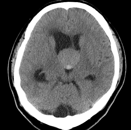
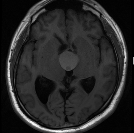
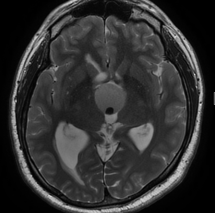
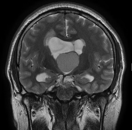
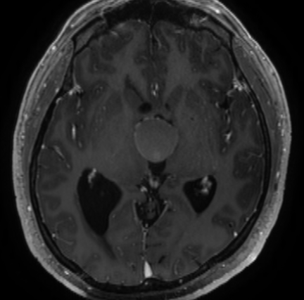
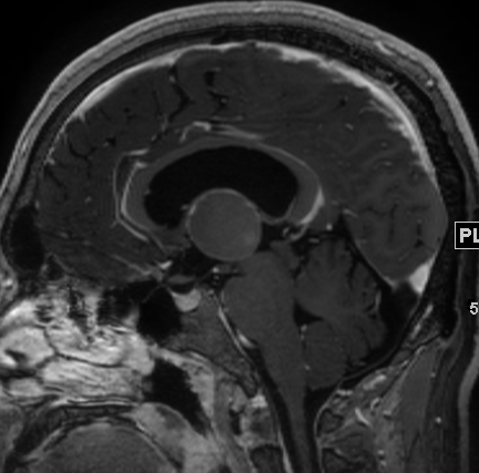
| Discussion | <문제해설:> Noncontrast CT scan shows isoattenuating mass-like lesion with small high attenuating nodule in the 3rd ventricle near the foramen of Monro causing hydrocephalus. This lesion demonstrates high signal intensity on T1WI and intermediate signal intensity on T2WI with T1 iso and T2 low signal intense nodule. Contrast-enhanced T1WI shows thin rim enhancement without solid enhancing component. Colloid cysts are unilocular, mucin-containing cysts that are almost always found wedged into the top of the third ventricle at the foramen of Monro. Colloid cysts are well-delineated round or ovoid masses. Imaging appearance depends on their viscosity and/or cholesterol content. The relative amounts of mucous material, cholesterol, protein, and water content all affect density/signal intensity. Desiccated, inspissated cysts appear very different from water-rich lesions. Density on NECT correlates directly with the hydration state of the cyst contents. Nearly two-thirds of all CCs are hyperdense compared to brain while one-third are iso- to hypodense. Signal intensity on T1WI reflects cholesterol concentration. Most CCs are hyperintense compared to brain, but one-third are isointense. CCs generally do not enhance. A thin peripheral rim of enhancement can be seen in some cases. |
| Correct answer |
Correct Answer
Semi-Correct Answer |