HOME > Weekly Case > Weekly Case
Weekly Case
종결
Case No. 414
2016-07-01~2016-07-31
출제자 : 안성수
Hit : 348
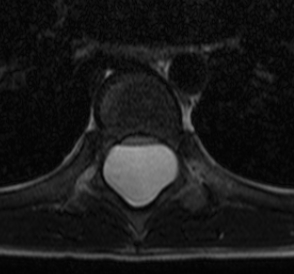
| Period | 2016-07-01~2016-07-31 |
|---|---|
| Diagnosis | Extradural arachnoid cyst |
| Clinical information | M/10 CC: Foot drop, gait disturbance |
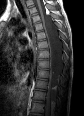
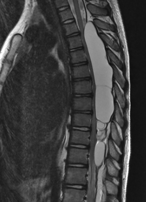
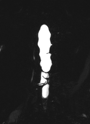
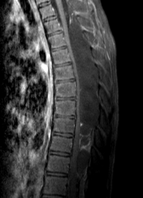
| Discussion | <문제해설> Multiloculated cystic lesion showing T1 low and T2 high signal intensity is seen in the extradural space of T3-T11 extending through the adjacent neural foramina. It causes dorsal compression and flattening of thecal sac. The lesion is associated with vertebral posterior body scalloping, suggesting longstanding mass effect. Spinal extradural arachnoid cysts are a rare cause of spinal cord compression. These cysts most commonly occur in the middle to lower thoracic spine (65%). Extradural arachnoid cysts develop from protrusions of arachnoid herniating through a small dural defect. The cysts have a pedicle in communication with the spinal subarachnoid space and, because of their origin, contain CSF. Magnetic resonance imaging is useful in detecting the mass, and the signal characteristics are consistent with those of CSF. Computed tomography myelography is better for detecting the dural defect when communication between the cyst and the subarachnoid space occurs and thus allows accurate diagnosis of these lesions. |
| Correct answer |
Correct Answer
Semi-Correct Answer |