HOME > Weekly Case > Weekly Case
Weekly Case
종결
Case No. 400
2016-02-01~2016-02-29
출제자 : 신나영
Hit : 448
| Period | 2016-02-01~2016-02-29 |
|---|---|
| Diagnosis | Metastatic mucinous adenocarcinoma |
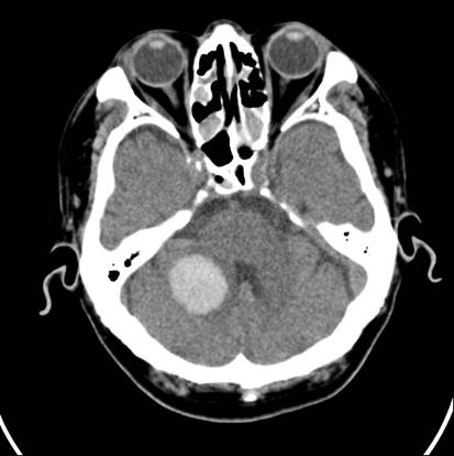
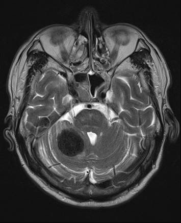
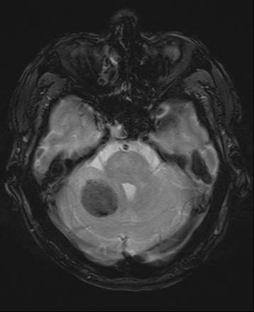
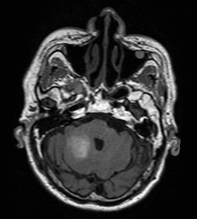
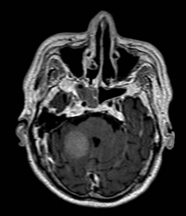
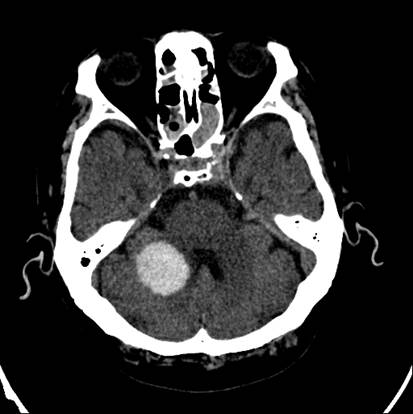
| Clinical information | M/70 C.C: dysarthria, left side weakness 마지막 사진은 3일 후 시행한 CT. |
| Discussion | Tip Hx: 1년전 대장암 수술 후 항암치료 중. Pathology 상 hemorrhage, calcification 없음. Brain metastasis from colorectal cancer is rare (7.6 %). Mucin-containing masses have often been reported to show low attenuation on CT. Due to its high water content, mucus has CT attenuation and MRI signal similar to that of water and does not enhance after injection of intravenous contrast medium; however, a chronic entrapped mucus collection can undergo a gradual transformation. The increased production of mucus protein is accompanied by a gradual reabsorption of water with a decrease in free water content and an increase in protein concentration. On CT, concentrated mucus may have attenuation values above 20 HU, and even as high as 130 HU. The value is similar to that of acute hematoma. The high viscosity of macromolecular material, such as mucin, causes shortening of the T1 and T2 relaxation time, resulting in T1 hyperintensity and T2 hypointensity. In this case, the observed no blooming artifact on GRE images was against a cerebellar hematoma. Also, subtle peripheral contrast enhancement on CE-T1WI and no change of high density lesion on serial CT scan suggested tumorous condition. Reference: J Korean Neurosurg Soc 34 : 252-253, 2003 Surg Neurol. 1999 Jul;52(1):46-9. http://www.eurorad.org/eurorad/case.php?id=1845 |
| Correct answer |
Correct AnswerSemi-Correct Answer |