HOME > Weekly Case > Weekly Case
Weekly Case
종결
Case No. 398
2016-02-01~2016-02-29
출제자 : 신나영
Hit : 373
| Period | 2016-02-01~2016-02-29 |
|---|---|
| Diagnosis | Methanol intoxication |
| Clinical information | F/27 C.C: N/V, dizziness, blurred vision 이후 Dyspnea x 1일 2개월 전부터 알코올로 세척된 제품을 검사하는 작업. |
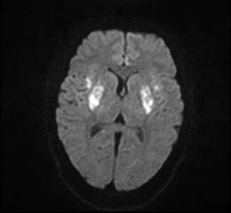
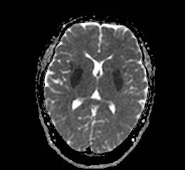
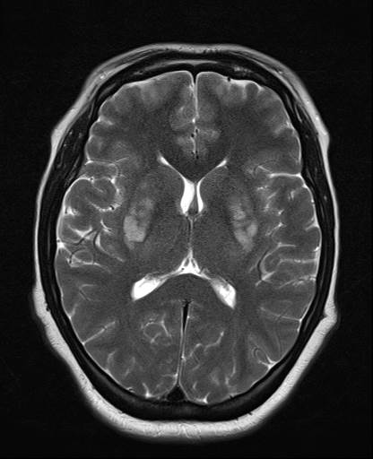
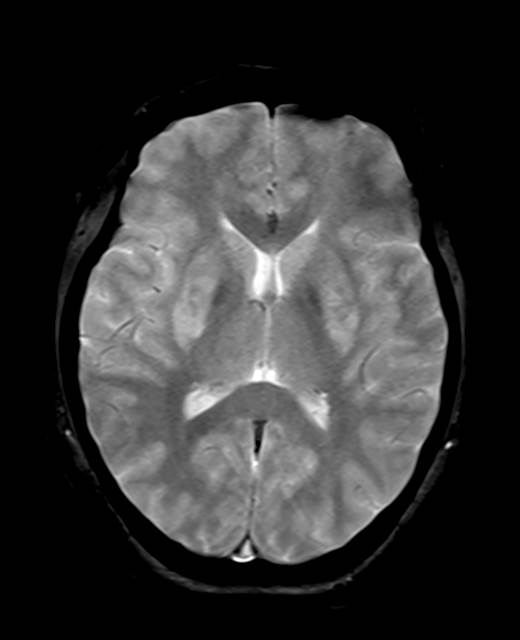
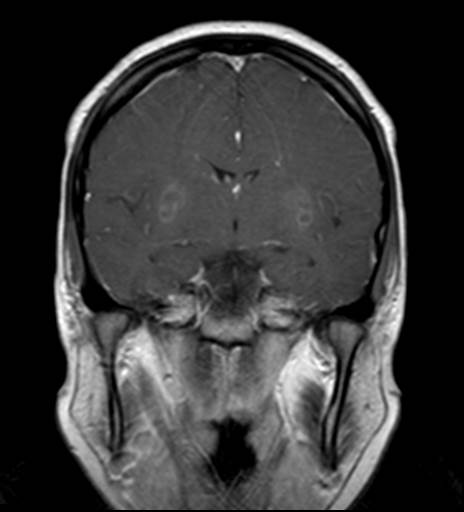
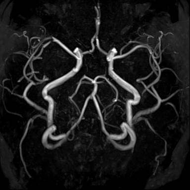
| Discussion | 1. Etiology - MtOH : a common component of solvents, varnishes, perfumes, paint removers, antifreeze, and gasoline mixtures. - It can be accidentally or intentionally ingested, inhaled, or absorbed transdermally. - MtOH metabolized to formaldehyde, formic acid, causing "anion gap acidosis" - Select toxic effect on putamen, optic nerves - Commercial products containing methanol include antifreeze, paint remover, photocopying fluid 2. Presentation - A peculiarity of MtOH poisoning is the latent period between ingestion and the appearance of clinical symptoms. Symptom onset is variable and often delayed, especially if ethanol is ingested simultaneously, as this slows MtOH metabolism. Between 85-90% of patients present with visual disturbances. Three-quarters of all patients have nonspecific gastrointestinal symptoms such as nausea and vomiting. Approximately 25% are comatose on admission. 3. Treatment - MtOH is effectively treated with alkali to combat acidosis, antidotes (ethanol or fomepizole) to block production of formic acid, and hemodialysis to remove MtOH and formate. 4. Imaging General Features - Best diagnostic clue: Bilateral hemorrhagic putaminal necrosis - Location: Putamen, hemispheric white matter, Caudate nuclei may be involved Uncommon: Corpus callosum, brainstem, cerebellum CT Findings - NECT: Bilateral hemorrhagic putaminal necrosis, White matter hypodensity may be seen MR Findings - T1WI : Bilateral hemorrhagic putaminal necrosis is characteristic Hemorrhagic subcortical necrosis White matter hypointense lesions (confluent hemispheric ± optic nerves) - T2WI: Lesions typically hyperintense Acute hemorrhagic necrosis may cause hypointense foci - FLAIR: Lesions all typically hyperintense - T2* GRE: Hypointense foci in putamina - DWI: Restriction (high signal) in putamina ± white matter - T1WI C+: May see subtle enhancement |
| Correct answer |
Correct Answer
Semi-Correct Answer |