HOME > Weekly Case > Weekly Case
Weekly Case
종결
Case No. 397
2016-01-01~2016-01-31
출제자 : 박지은
Hit : 423
| Period | 2016-01-01~2016-01-31 |
|---|---|
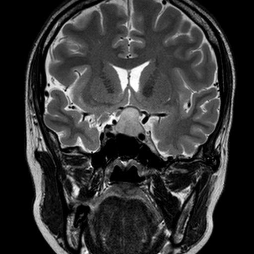
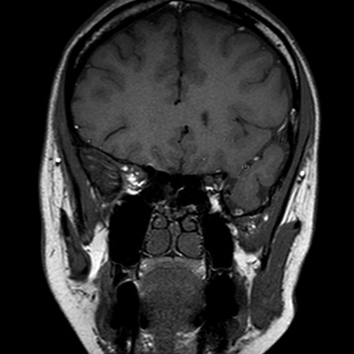
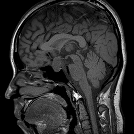
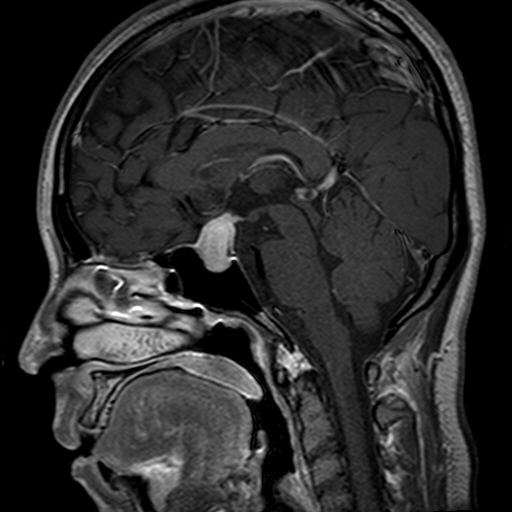
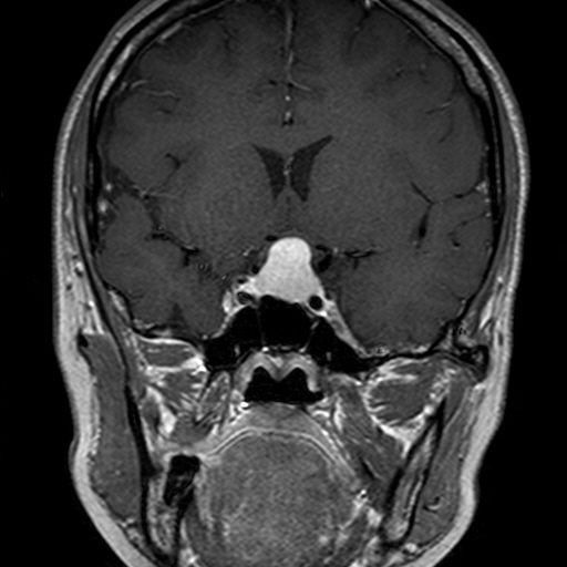
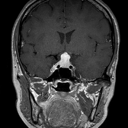
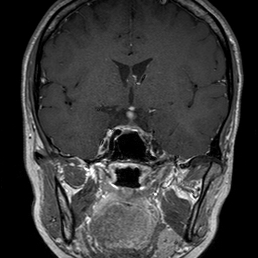
| Diagnosis | lymphocytic hypophysitis |
| Clinical information | 28/F CC: 임신 29주, 1달전부터 시작된 시력 저하 |
| Discussion | Autoimmune inflammatory disorder of the pituitary gland Demographics women of child bearing age, usually late in pregnancy or shortly after childbirth Reestablished immune competence in the late pregnancy/peripartum period Clinical Manifestation : Headache, diabetes insipidus, ACTH deficit, hyperprolactinemia. Imaging findings Thickened and nontapering stalk Symmetrically enlarged pituitary gland Loss of posterior pituitary bright spot Adjacent dural or sphenoid sinus mucosal thickening Hypophysitis / Pituitary adenoma Age - Younger / Older Diabetes insipidus-Common / Rare Pituitary expansion-Symmetric/ Asymmetric Posterior “bright spot”-Loss / Preserved Stalk -Enlarged, nontapering / Displaced Signal- Homogeneous / Heterogeneous Enhancement- Strong / Moderate Reference 1. Gutenberg A, Larsen J, Lupi I, et al. A radiologic score to distinguish autoimmune hypophysitis from nonsecreting pituitaary adenoma preoperatively. Am J Neuroradiol. 2009; 30:1766-1772. 2. Cheng CC. Ezzat S, Smyth HS, et al. The spectrum and significance of primary hypophysitis. JCEM. 2001; 86(3):1048-1053. 3. Osborn’s brain : Imaging, pathology, and anatomy, Osborn AG, et al. Amirsys, 2013, 713-716. |
| Correct answer |
Correct Answer
Semi-Correct Answer |