HOME > Weekly Case > Weekly Case
Weekly Case
종결
Case No. 396
2016-01-01~2016-01-31
출제자 : 박지은
Hit : 379
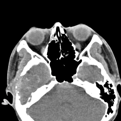
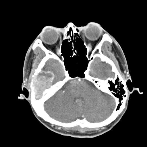
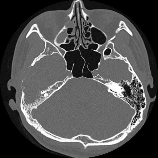
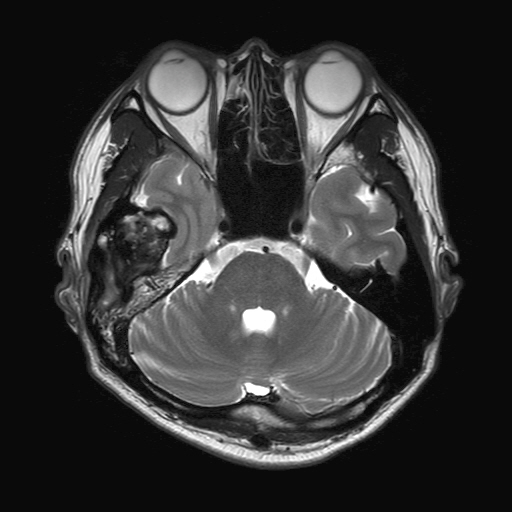
| Period | 2016-01-01~2016-01-31 |
|---|---|
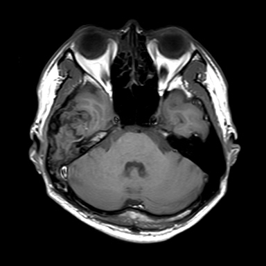
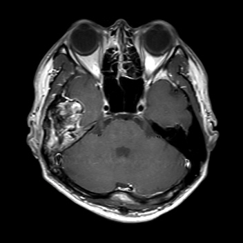
| Diagnosis | Giant cell tumor, right temporal bone |
| Clinical information | M/35 C.C : Right otalgia, hearing disturbance |
| Discussion | [Skull base Giant cell tumor (GCT)] Benign neoplasm but often exhibits aggressive behavior Demographics Age : Peak incidence 3rd-4th decade Gender : Female predominance Location Mainly arises from the epiphyses of long bones (m/c distal femur) Rarely arises from skull GCT (temporal, sphenoid bone) Imaging findings CT NECT Soft tissue algorithm : mildly hyperdense soft tissue mass Bone algorithm : overlying thinned cortical shell often sclerotic, focally interrupted CECT Marked homogeneous or heterogeneous enhancement MR T1WI Mixed iso- to hyperintense to gray matter Hypointense rim commonly seen T2WI Mixed signal, predominantly iso- to slightly hyperintense to gray matter Occasionally diffusely hypointense (hemosiderin, calcification) Markedly hypointense rim Enhanced T1WI Marked homogneous or heterogeneous enhancement Reference 1. Diagnostic imaging Head and Neck, Giant cell tumor, skull base I 3 50-53 2. Byun JH, Park KB, Ko JS, Ahn SK. Giant cell tumor of infratemporal fossa and mandibular doncyle: A case report Int Adv Otol 2015; 11(1):88-91 |
| Correct answer |
Correct Answer
Semi-Correct Answer |