HOME > Weekly Case > Weekly Case
Weekly Case
종결
Case No. 386
2015-10-01~2015-10-31
출제자 : 차지훈
Hit : 320
| Period | 2015-10-01~2015-10-31 |
|---|---|
| Diagnosis | Granulomatosis with polyangiitis |
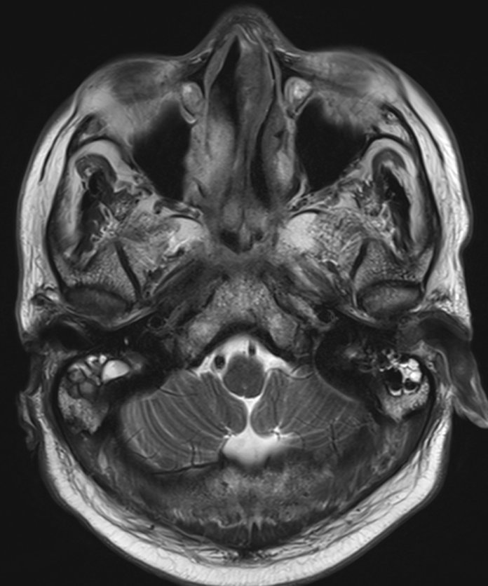
| Clinical information | M/62 C.C> headache |
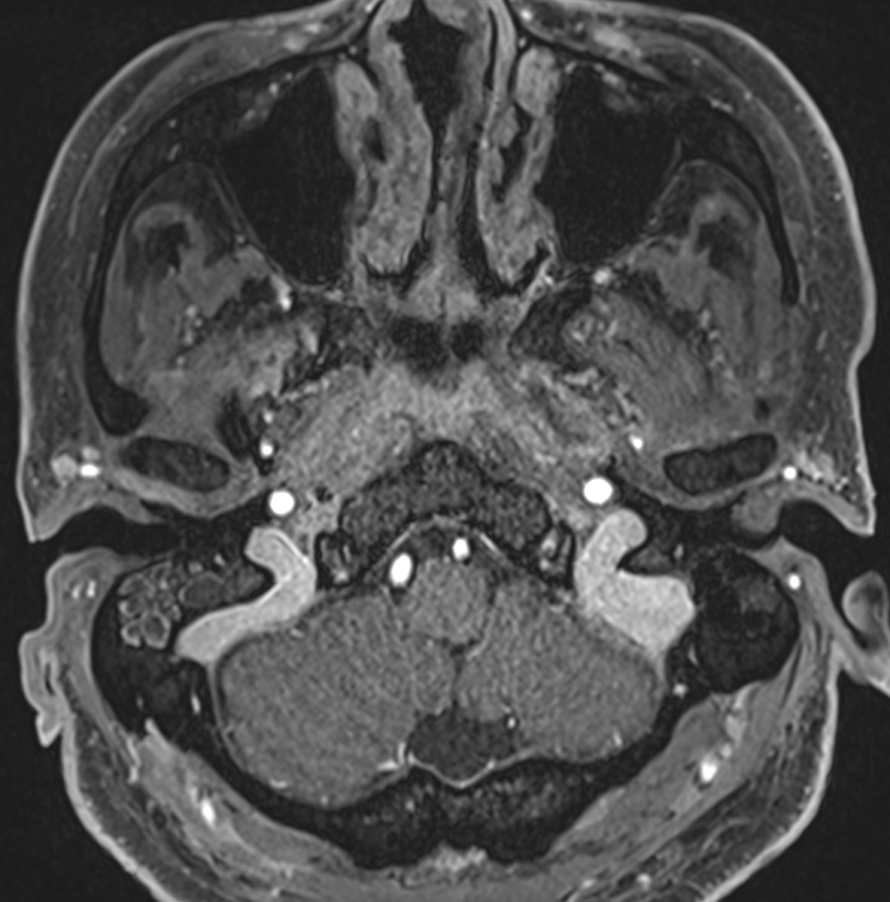
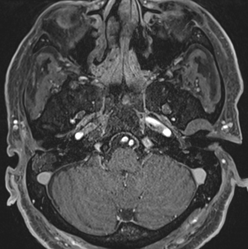
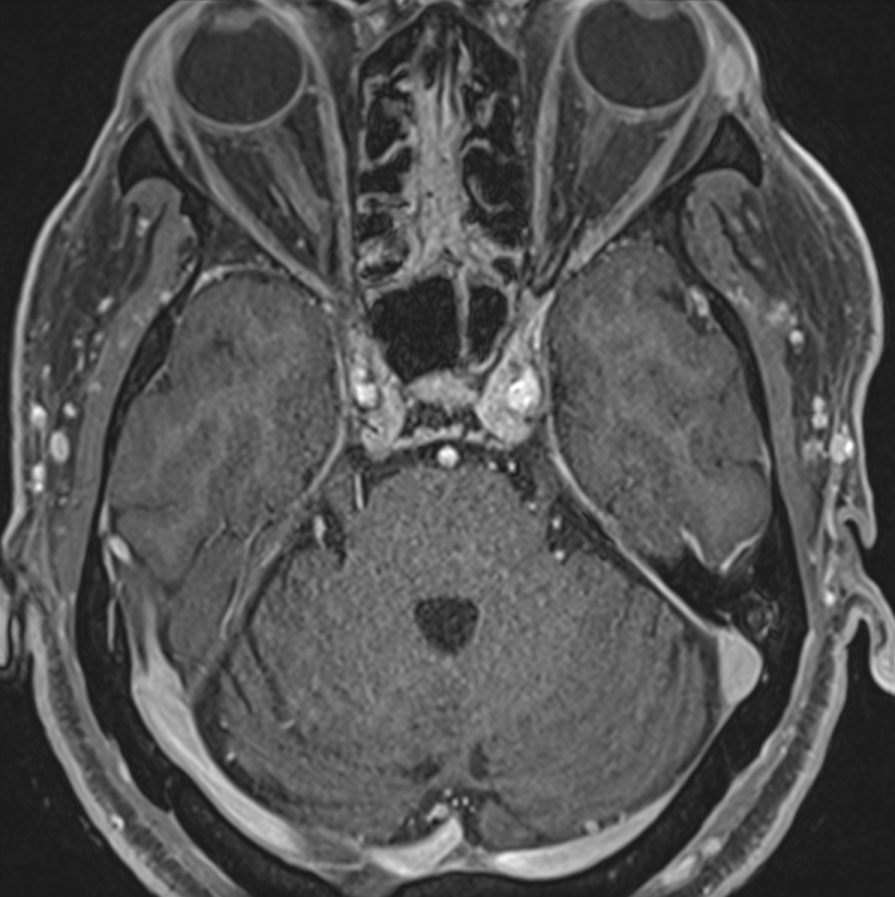
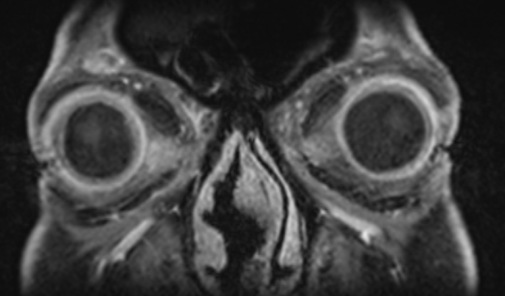
| Discussion | Imaging findings - Enhancing soft tissue lesion involving nasopharynx, eustachian tube with bilateral middle ear effusion. - Enhancing thickening of both anterior sclera - Nasal septal thickening with central nonenhancing necrotic area with T2 low signal intensity Review - Name changed: Wegener's granulomatosis (WG) ANCA-associated granulomatous vasculitis (2006) granulomatosis with polyangiitis (GPA) (ACR, 2011) - Autoimmune necrotizing granulomatous vasculitis involving Small and medium sized vessels involving upper & lower respiratory tract, kidney, skin, joint H&N involvement in 72-100% of patients 40-60 years & M > F Positive cANCA (cytoplasmic anti-neutrophil cytoplasmic antibody, anti-proteinase 3 (PR3)), 85-98% specificity, 10-50% may negative - Imaging Nodular soft tissue (may hypointense on T2WI compared to inflammed mucosa) in nose with mucosal ulceration and necrosis Nasal septal perforation (lead to saddle nose deformity), turbinate/lateral nasal wall destruction associated with chronic sinusitis, sclerotic to destructive sinus wall change, periantral soft tissue infiltration Orbital invasion including scleritis, conjunctivitis, uveitis: most common extrasinonasal H&N site Nasopharynx and eustachian tube involvement mimics malignancy |
| Correct answer |
Correct Answer
Semi-Correct Answer |