HOME > Weekly Case > Weekly Case
Weekly Case
종결
Case No. 380
2015-08-01~2015-08-31
출제자 : 이경미
Hit : 378
| Period | 2015-08-01~2015-08-31 |
|---|---|
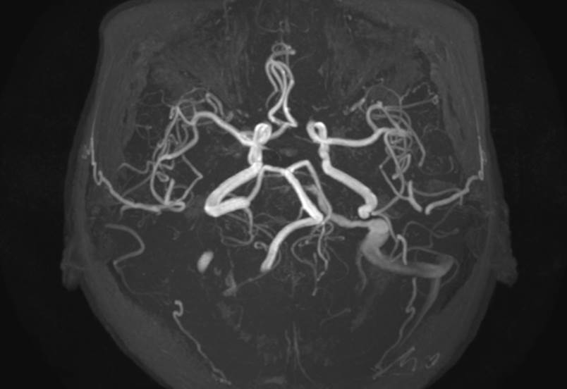
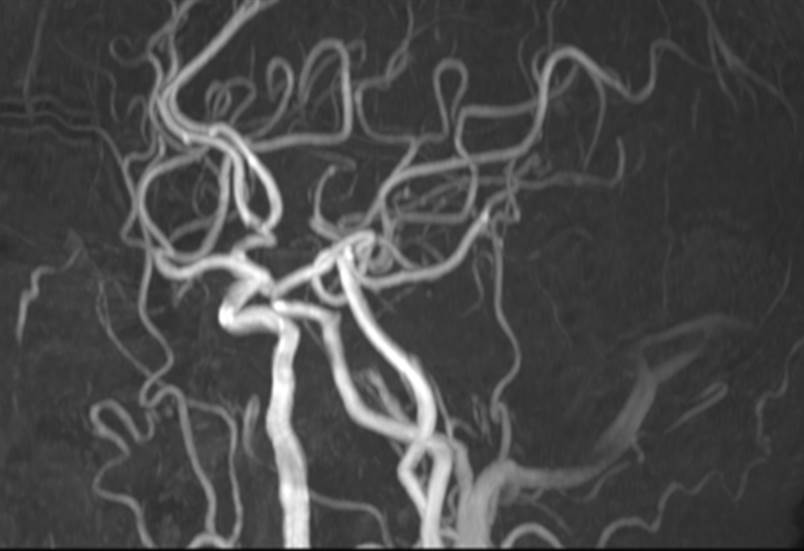
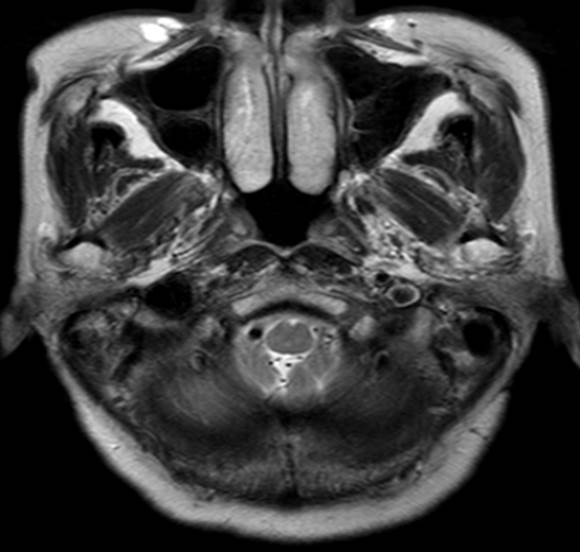
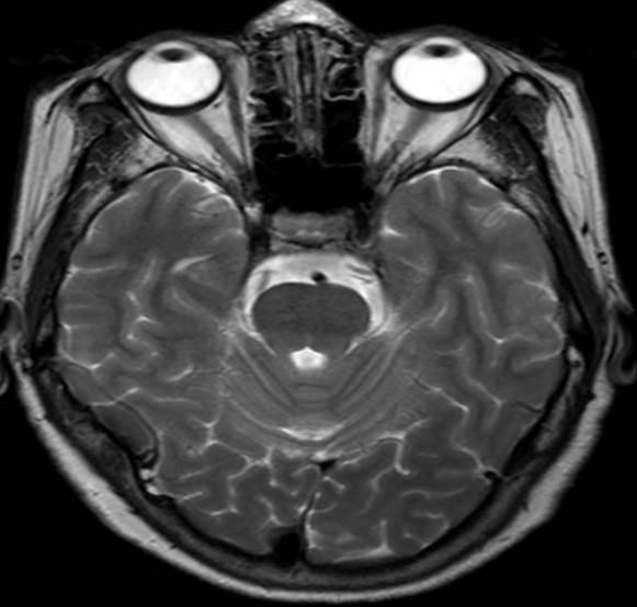
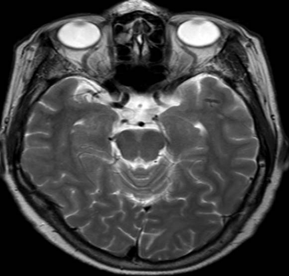
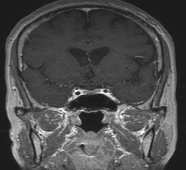
| Diagnosis | Jugular venous reflux |
| Clinical information | M/61 CI> 건강검진 |
| Discussion | JVR is known to be caused by physiologic compression of the brachiocephalic vein that leads to stagnation or reversal of the internal jugular vein flow. Although most reported cases of JVR were detected on the left side using MRI and were caused by compression of the left brachiocephalic vein between the aorta and sternum. JVR might be related to some neurologic diseases or symptoms, such as transient global amnesia and transient monocular blindness. JVR vs. cavernous dural AVF 1. JVR signal was predominantly on the left side in contrast to cavernous DAVF. JVR mainly involved the inferior petrosal sinus, sigmoid sinus, or transverse sinus without involvement of the cavernous sinus, whereas cavernous DAVF always involved the cavernous sinus. 2. CE-MRA were divided into subgroups representing the arterial phase and the arteriovenous phase (i.e., venous opacification group). The presence or absence of early opacification of the cavernous sinus in the arterial phase of CE-MRA appeared to be an imaging finding for distinguishing between JVR and cavernous DAVF. 3. All patients with JVR had contralateral jugular vein drainage, whereas cavernous DAVF had ipsilateral or bilateral jugular drainage. It seems that the ipsilateral jugular vein in JVR has some degree of resistance, due to the retrograde flow and elevated venous pressure, so the intracranial venous blood drains more easily through the contralateral jugular vein where the venous pressure is low. 4. flow-void cluster, exophthalmos, engorgement of the superior ophthalmic vein, or enlargement of the cavernous sinus; the lack of these findings can also be helpful for establishing the diagnosis of JVR. |
| Correct answer |
Correct Answer
Semi-Correct Answer |