HOME > Weekly Case > Weekly Case
Weekly Case
종결
Case No. 378
2015-07-01~2015-07-31
출제자 : 안성수
Hit : 363
| Period | 2015-07-01~2015-07-31 |
|---|---|
| Diagnosis | Hemangiopericytoma |
| Clinical information | F/30 C.C> Headache |
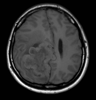
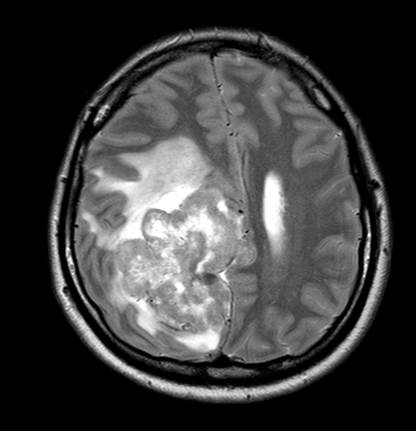
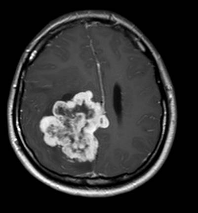
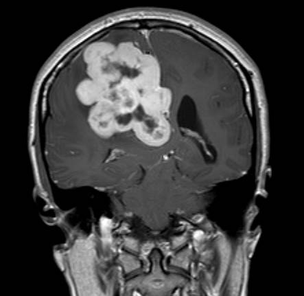
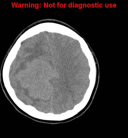
| Discussion | 해설: Axial T1- and T2WI show lobulated mass attached at posterior falx with peritumoral edema. Dural tail is not seen on axial and coronal postcontrast T1WI. There is no calcification within the mass on CT. Lobular enhancing extraaxial mass with dural attachment, ± skull erosion May mimic meningioma, but without Ca++ or hyperostosis CT: iso- to hyperdense and often heterogeneous, calvarial erosion may be seen MRI: similar signal intensity to meningioma, often heterogeneous |
| Correct answer |
Correct Answer
Semi-Correct Answer |