HOME > Weekly Case > Weekly Case
Weekly Case
종결
Case No. 372
2015-05-01~2015-05-31
출제자 : 이아름
Hit : 352
| Period | 2015-05-01~2015-05-31 |
|---|---|
| Diagnosis | Sturge Weber syndrome |
| Clinical information | F/14 C.C.: headache, N/V, right visual disturbance left side paresthesia 진단은? |
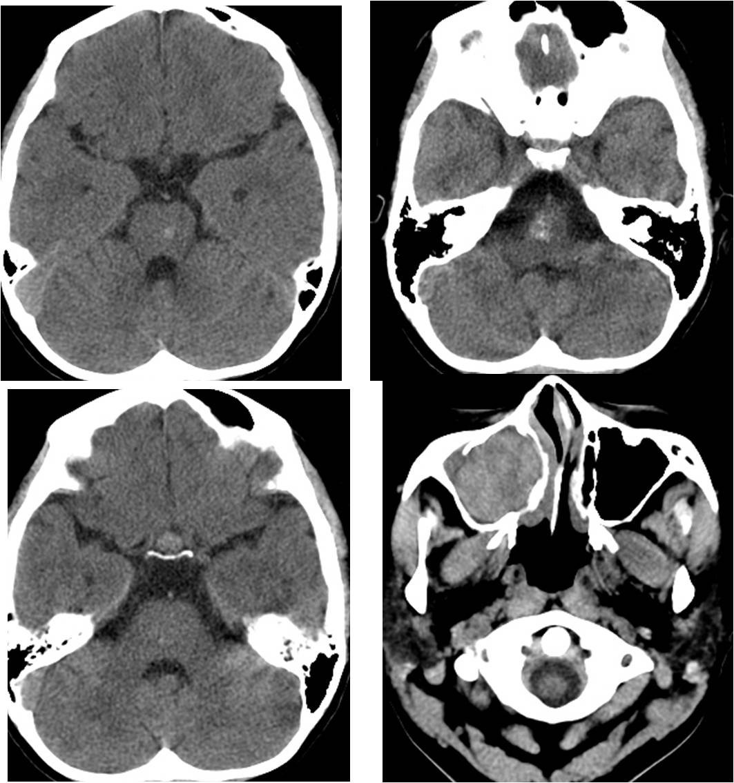
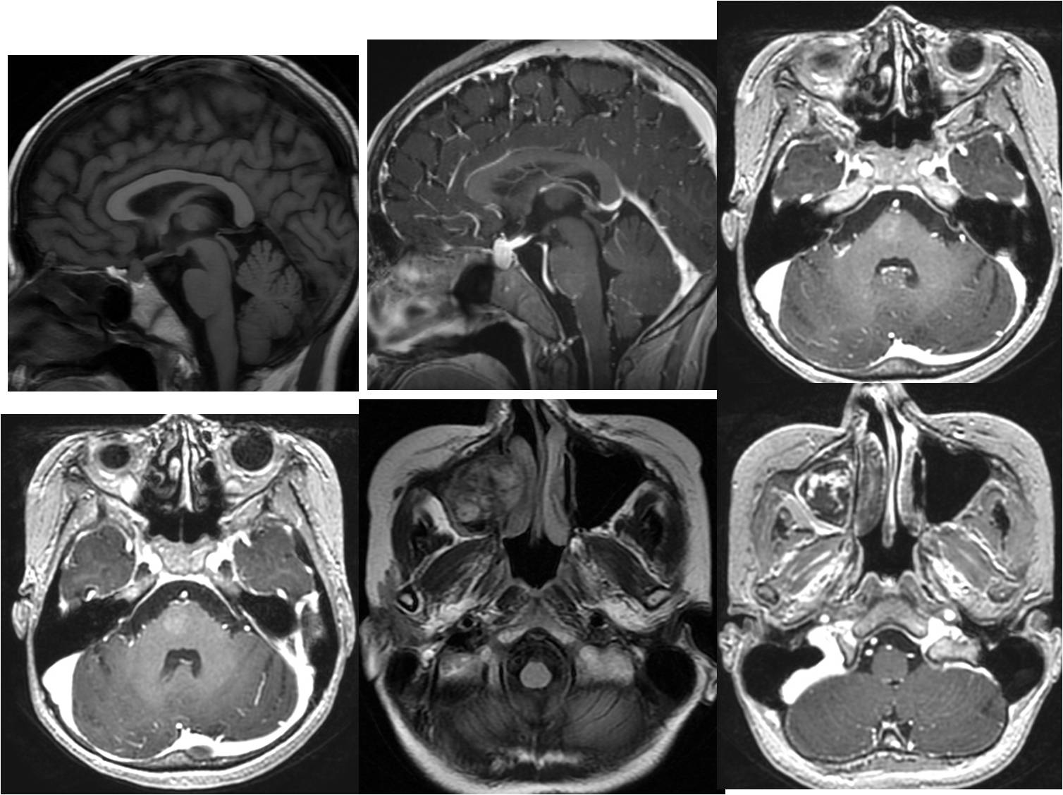
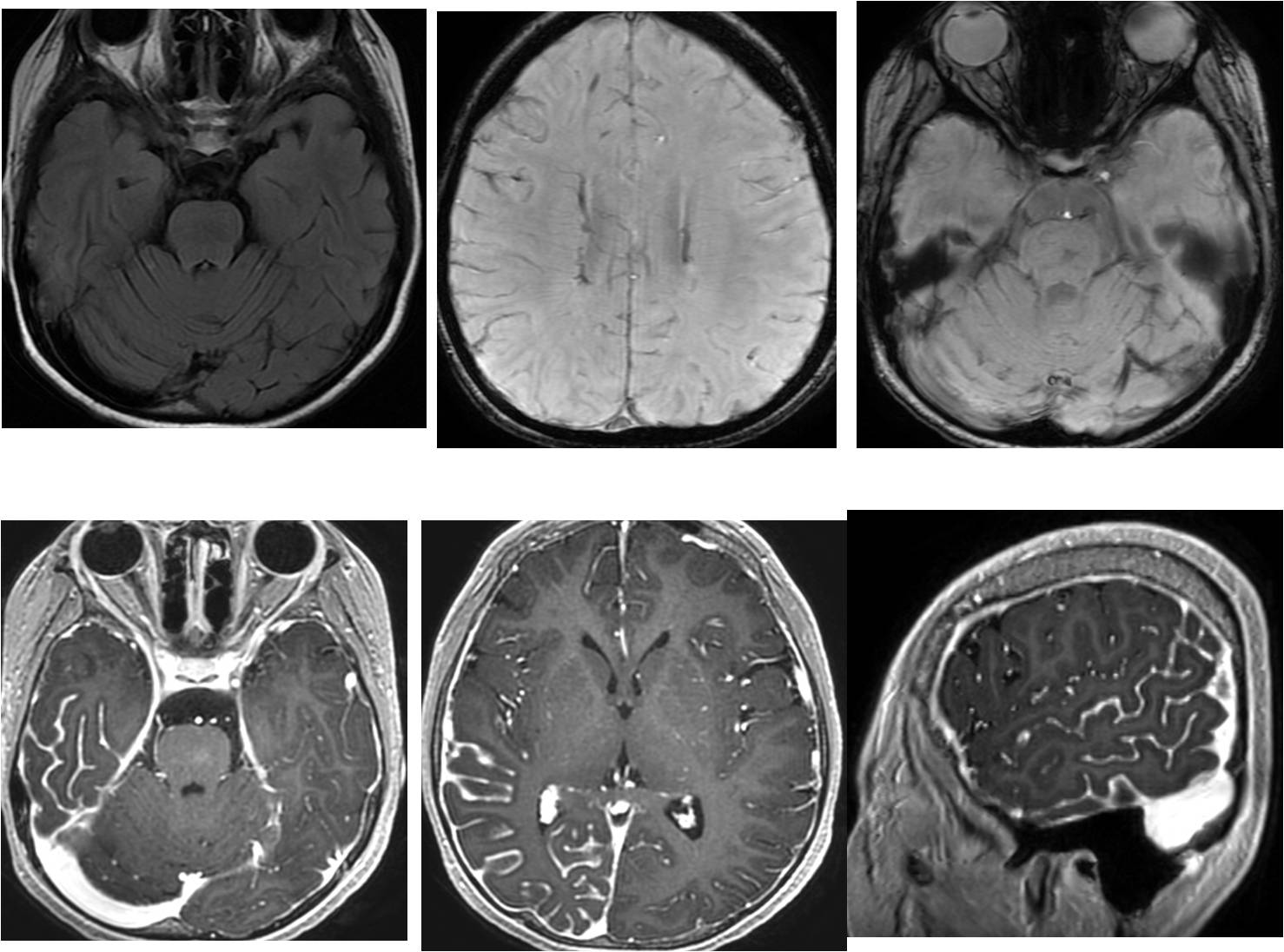
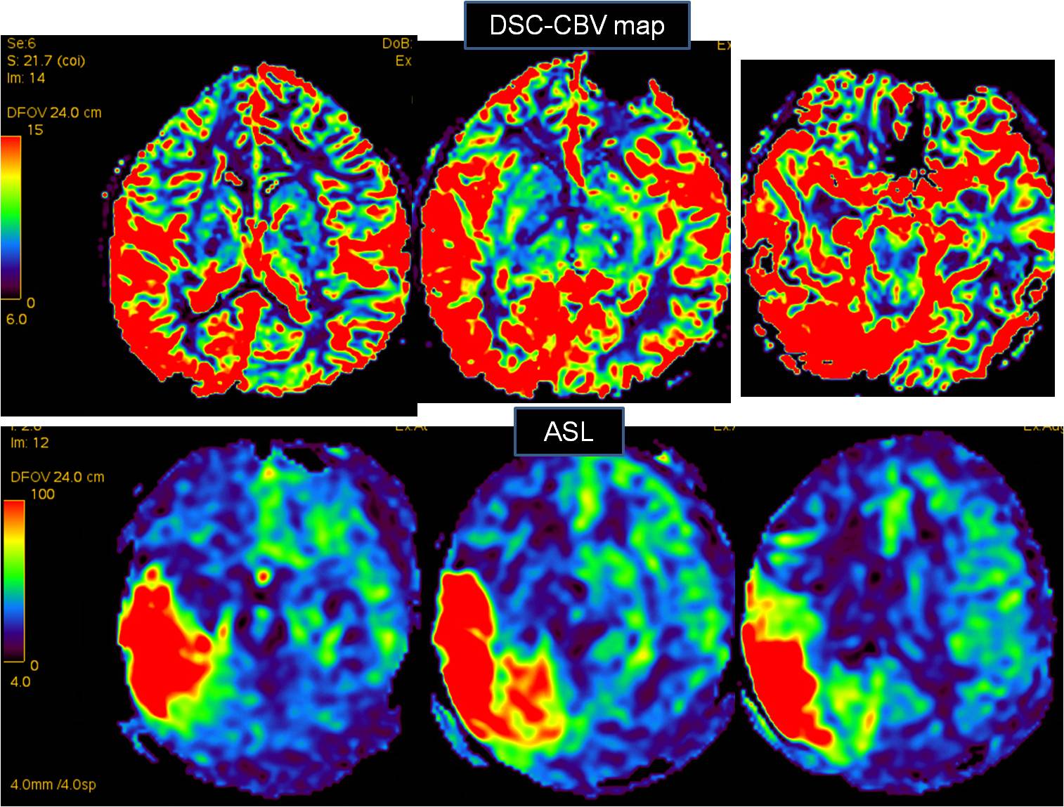
| Discussion | 정답: Sturge Weber syndrome 해설: Imaging findings (1-4) and Clinical finding (5) 1. Leptomeningeal angiomatosis and choroidal angioma in Rt. Lateral ventricle 2. Capillary telangiectasia in brain stemBehcet disease (Neurobehcet) 3. High attenuated, expansile enhancing mass in Rt. maxillary sinus : Cavernous hemangioma with organizing hematoma 4. Pituitary hyperplasia 5. Clinical finding : Congenital glaucoma history Multiple whole body and facial nevus flammeus and capillary hemangioma C/W Sturge Weber disease Sporadic congenital neurocutaneous disorder Meningeal tangle of vessels à angioma (pia matter) Fetal cortical veins fail to develop normally Angiomatosis involving face, choroid of eye and leptomeninges à Imaging features: sequelae of progressive venous occlusion and chronic venous ischemia Gender: M = F Three types of SWS Type I : facial and leptomeningeal angioma associated with possible glaucoma Type II : facial angioma Type III : exclusive endocranial involvement |
| Correct answer |
Correct Answer
Semi-Correct Answer |