HOME > Weekly Case > Weekly Case
Weekly Case
종결
Case No. 371
2015-05-01~2015-05-31
출제자 : 이아름
Hit : 351
| Period | 2015-05-01~2015-05-31 |
|---|---|
| Diagnosis | Neurofibromatosis type I |
| Clinical information | 21/M C.C : health check up 진단은? |
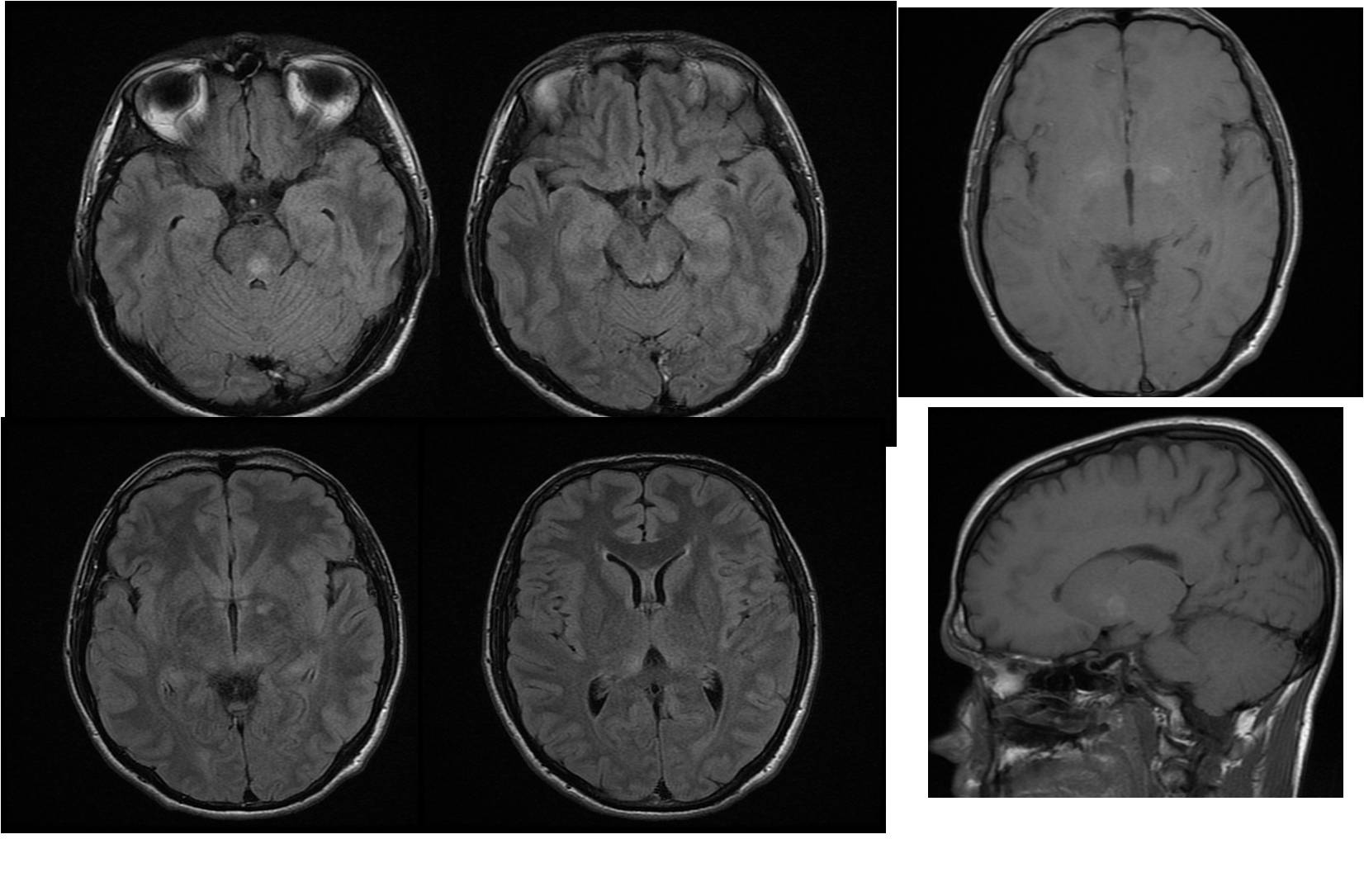
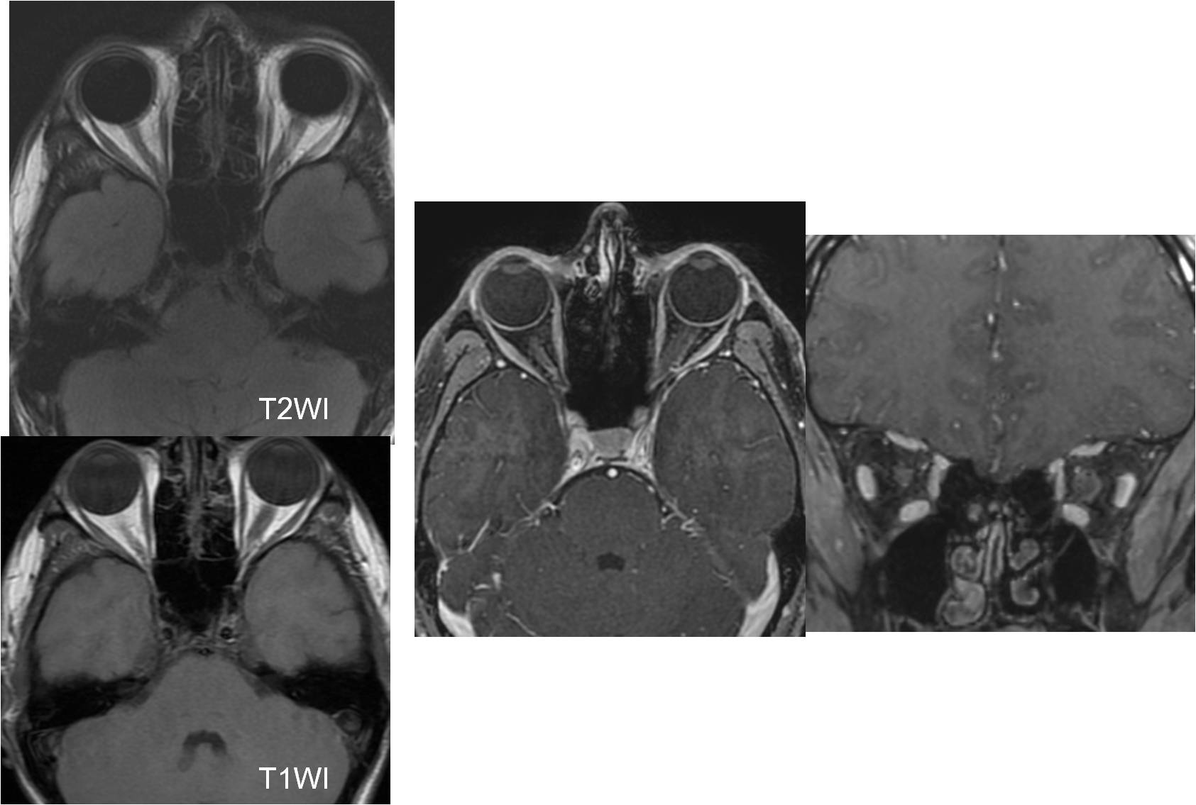
| Discussion | 정답: Neurofibromatosis type I 해설: Imaging findings • Bilateral symmetrical T1 high SI lesions in the both globus pallidus and small T2 high SI lesions in left BG and pons: almost disappeared on FU MRI • Slight enlargement of the left optic nerve with focal T2 high intensity, suggesting either optic neuritis or optic nerve glioma • 1) Non-neoplastic CNS lesions in the NF-I : Dysplastic WM lesions : Globus pallidus, thalamus, brainstem, cerebellar WM, centrum semiovale T2/FLAIR : multifocal, high SI T1WI : Iso SI but GP lesions are often mild high SI Increase in size and number until 10 years old Wane and resolve, rare in adult Smaller than 2cm, little or no mass effect No enhancement 2) CNS neoplasm : Optic nerve glioma Optic pathway pilocytic astrocytoma (M/C) Diffuse fusiform enlargement of one or both optic nerves May extend into optic chiasm, hypothalamus, temporal bone, brainstem Iso SI on T1WI, iso to moderate high SI on T2WI Enhancement: variable, none to striking |
| Correct answer |
Correct Answer
Semi-Correct Answer |