HOME > Weekly Case > Weekly Case
Weekly Case
종결
Case No. 367
2015-03-02~2015-03-31
출제자 : 정원상
Hit : 366



| Period | 2015-03-02~2015-03-31 |
|---|---|
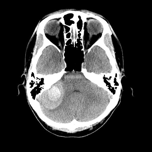
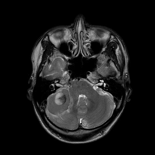
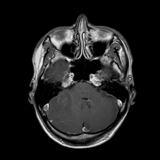
| Diagnosis | Acute hemorrhage of underlying cavernous malformation |
| Clinical information | M/43 C/C headache (3 days ago) |
| Discussion | Acute hemorrhage of underlying cavernous malformation Cerebral cavernous venous malformations (commonly known as cavernous haemangioma) are common cerebral vascular malformations, usually with characteristic appearances on MRI. Many alternative terms have been used including cavernous hemangioma, cerebral cavernous malformation or simply cavernoma. The majority of lesions remain asymptomatic throughout life and are found incidentally. Presentation due to haemorrhage is with a seizure or focal neurological deficit. The risk of haemorrhage is 1% per year for familial cases, and somewhat less for sporadic lesions. Radiographic features CT If large then a region of hyperdensity can be seen. If there has been a recent bleed then it is more conspicuous and may be surrounded by a mantle of edema. MRI MRI is the modality of choice, demonstrating a characteristic “popcorn” or "berry" appearance with a rim of signal loss due to hemosiderin, which demonstrates prominent blooming on susceptibility weighted sequences. T1 and T2 signal is varied internally depending on the age of the blood produces and small fluid fluid levels may be evident. Gradient Echo or T2* sequences are able to delineate these lesions better than T1 or T2 weighted images. In patients with familial or multiple cavernous angiomas GRE T2* sequences are very important in identifying the number of lesions missed by conventional Spin echo sequences. Susceptibility imaging (SWI) may have sensitivity equal to that of GRE in detecting these capillary telagiectasiae in brain. If a recent bleed has occurred then surrounding edema may be present. The lesions generally do not enhance Angiography / DSA Cavernous malformations are angiographically occult and do not demonstrate arteriovenous shunting. |
| Correct answer |
Correct Answer
Semi-Correct Answer |