HOME > Weekly Case > Weekly Case
Weekly Case
종결
Case No. 366
2015-03-02~2015-03-31
출제자 : 정원상
Hit : 360



| Period | 2015-03-02~2015-03-31 |
|---|---|
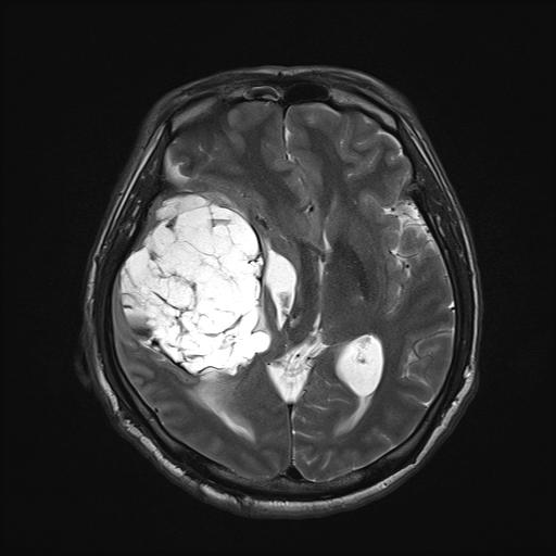
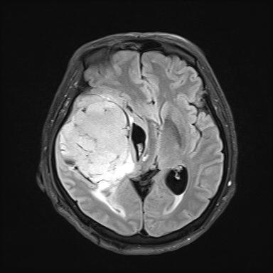
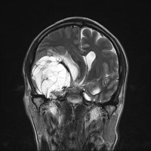
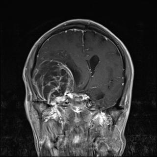
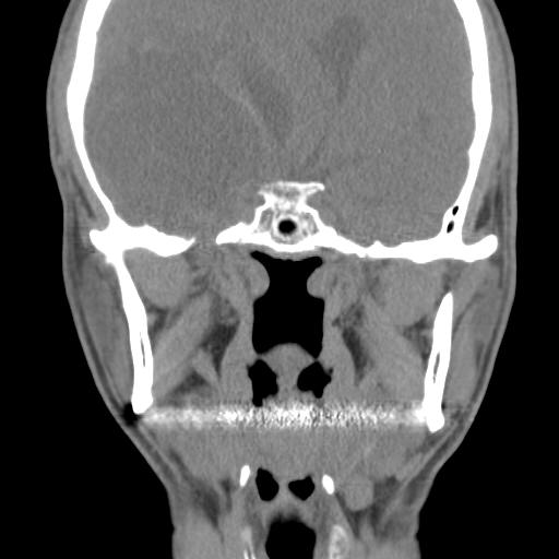
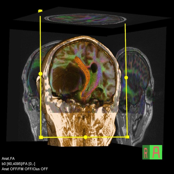
| Diagnosis | Intracranial schwannoma |
| Clinical information | 55/M C/C: dradual change of decreased visual field, decreased memory function |
| Discussion | Intracranial schwannoma Intracranial schwannomas (also referred to as neurinomas) are common benign tumours, accounting for 6-8% of all intracranial tumours. Schwannomas are most frequently encountered in middle aged and elderly adults with a predilection for females (F:M = 1.5-2:1), although they are found in all age groups. Radiographic features Although the appearance of individual schwannomas will be influenced by the nerve of origin and surrounding anatomy, they naturally share similar imaging characteristics. CT Schwannomas are typically isodense to brain, and can be difficult to identify, depending on location. Cystic areas (usually found in larger tumours) are hypodense, similar to CSF. Following administration of contrast they moderately enhance, often heterogeneously due to cystic areas. Bone algorithm is excellent at assessing bony margins, especially useful when the tumour extends into or thorough a foramen. As these lesions are slow growing they remodel the bone, with smooth borders. MRI MRI is the investigation of choice for assessment of intracranial schwannomas, not only due to greater contrast resolution, but also exquisite anatomical details, which allows for precise localisation of the tumour. T1 typically isointense to brain cystic areas, if present, are hypointense T2 typically somewhat hyperintense to brain cystic areas are hyperintense T1 C+ prominent enhancement |
| Correct answer |
Correct AnswerSemi-Correct Answer |