HOME > Weekly Case > Weekly Case
Weekly Case
종결
Case No. 365
2015-03-02~2015-03-31
출제자 : 정원상
Hit : 314

| Period | 2015-03-02~2015-03-31 |
|---|---|
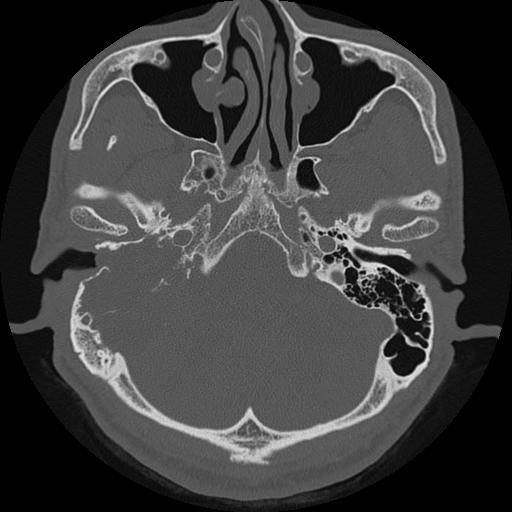
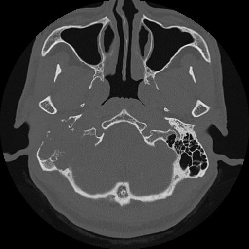
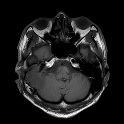
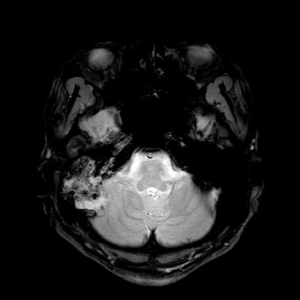
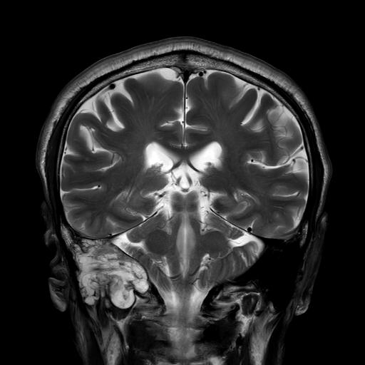
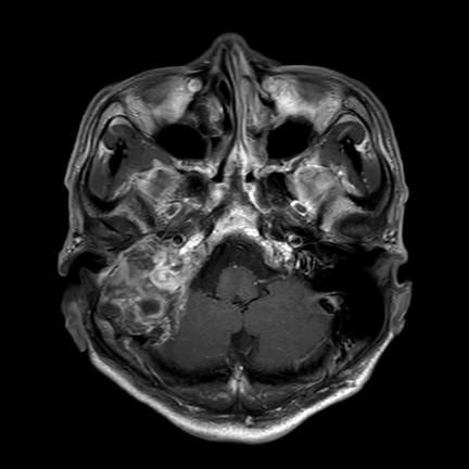
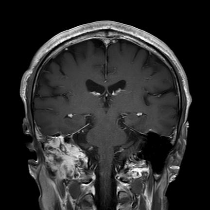
| Diagnosis | Facial nerve schwannoma |
| Clinical information | 58/M C/C hearing impairment (6months ago) |
| Discussion | Radiogrphic findings Same as that of schwannomas elsewhere, homogenous enhancement on both CT and MRI when small; heterogenous when large. CT In all cases, high resolution thin section bone CT of the temporal bone is required to assess the otic capsule. Bony scalloping and remodelling may be present. MRI Signal characteristics include T1: typically iso- to hypo-intense relative to gray matter T2: typically hyperintense; large lesion may show hetrogenous signal T1 C+: usually demonstrate homogeneous enhancement with larger lesions with cystic degeneration seen as focal intramural low signal intensity Differential diagnosis Depends on location on lesion along the facial nerve: when in an intraparotid location consider differential for a parotid mass. when at the cerebellopontine angle consider differential for cerebellopontine angle mass Reference Wiggins RH, Harnsberger HR, Salzman KL et-al. The many faces of facial nerve schwannoma. AJNR Am J Neuroradiol. 2006;27 (3): 694-9 |
| Correct answer |
Correct AnswerSemi-Correct Answer |