HOME > Weekly Case > Weekly Case
Weekly Case
종결
Case No. 363
2015-02-02~2015-02-28
출제자 : 최진우
Hit : 329
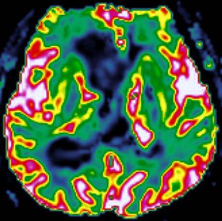
| Period | 2015-02-02~2015-02-28 |
|---|---|
| Diagnosis | Subependymoma |
| Clinical information | M/34 C.C: headache, vomiting (Onset: 5DA) |
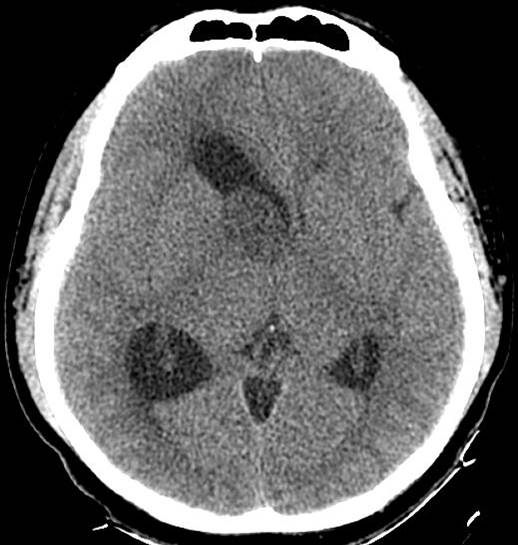
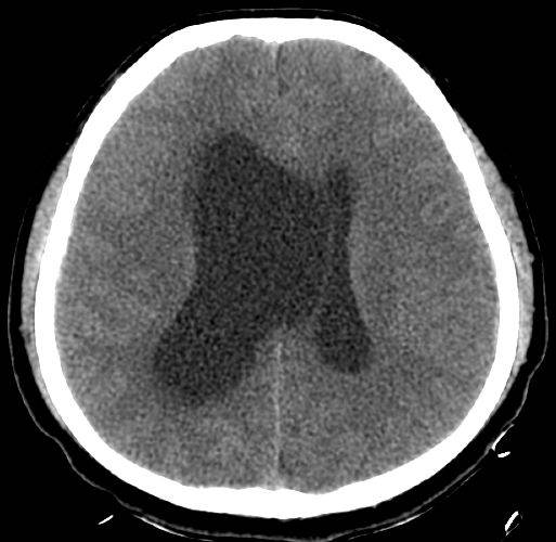
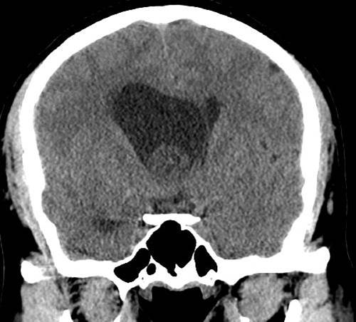
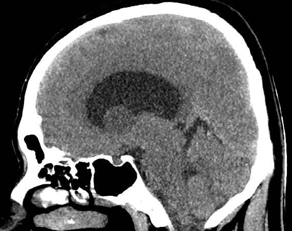
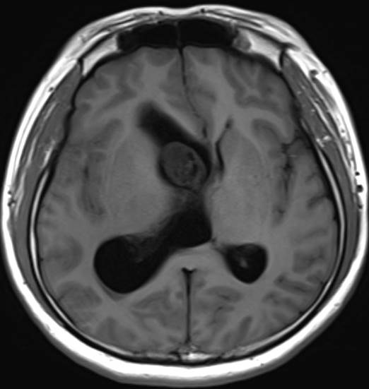
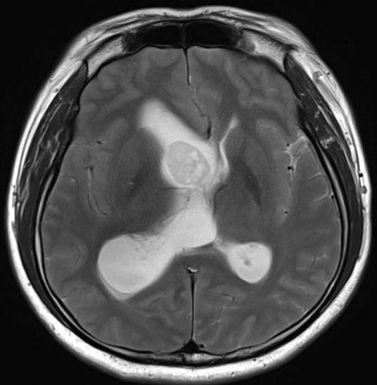
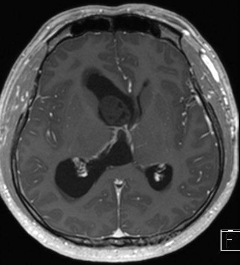
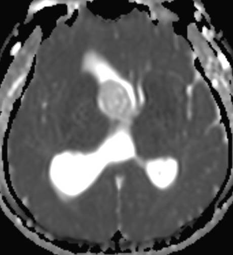
| Discussion | Subependymoma • Rare benign CNS tumor, may represent a transitional form between ependymomas and astrocytomas. (arise from the subependymal glial layer) • WHO grade I • >15 years (82%); in middle-aged and elderly adults • male predominance (male-to-female ratio, 2.3:1) • Lateral recesses of 4th vent.(50-60%), lateral vent.(30-40%), spinal cord • Asymptomatic(60%) • Gross total resection is curable!! • CT • Well-circumscribed, lobulated intraventricular mass • Predominantly iso to slightly hypoattenuation • Enhancement (-) • Calcification(30%), cystic degeneration(20%) • MRI • Isointense on T1WI and hyperintense on T2WI/FLAIR • Variable enhancement pattern, typically none to mild • Extension beyond the ventricular margins is rare • No edema seen in adjacent brain parenchyma References 1. Cerebral Intraventricular Neoplasms: Radiologic-Pathologic Correlation RadioGraphics 2002; 22:1473–1505 2. Intraventricular Neoplasms: Radiologic-Pathologic Correlation RadioGraphics 2013; 33:21–43 |
| Correct answer |
Correct Answer
Semi-Correct Answer |