HOME > Weekly Case > Weekly Case
Weekly Case
종결
Case No. 361
2015-01-01~2015-01-31
출제자 : 임현경
Hit : 417
| Period | 2015-01-01~2015-01-31 |
|---|---|
| Diagnosis | Japanese encephalitis |
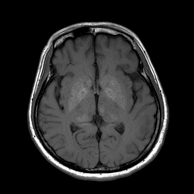
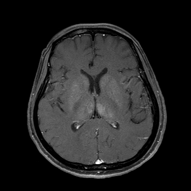
| Clinical information | Fever 및 mental change를 동반한 62세 여자. 가장 적합한 진단명은? |
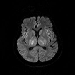
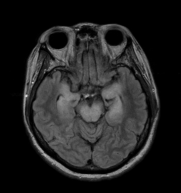
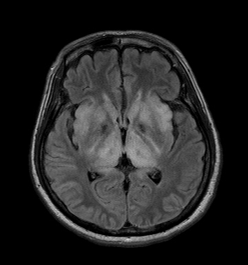
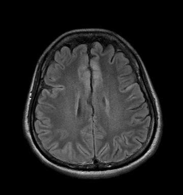
| Discussion | Etiology Japanese encephalitis (JE) is a mosquito-borne flaviviral encephalitis that remains a major health problem in countries in the Indian subcontinent, Southeast Asia, and the Far East. Forty-five thousand to 50,000 new cases occur annually. It occur in the summer rainy season, when conditions are ideal for mosquito breeding. Case fatality ranges from 10% to 60%. Up to 50% of those who recover are left with disabling neurologic deficits. Imaging Finding The MR imaging and CT findings of JE have been described as bilateral thalamic, substantia nigra, basal ganglia, brain stem, cerebellum, cerebral cortical, and white matter lesions. Bilateral T2 hyperintense and T1 hypointense to isointense thalamic lesions, especially hemorrhagic have been described as characteristic in an appropriate clinical setting. Hemorrhagic transformations have also been described in JE lesions, with corresponding expected T1 and T2 changes. The temporal lobe involvement pattern is fairly characteristic and mostly involves the hippocampus, usually sparing the rest of the temporal lobe. Differential considerations based on the MRI appearance are somewhat broad, including but not limited to primary viral encephalitis, acute encephalopathy, limbic encephalitis, and acute disseminated encephalomyelitis. Reference: 1. Abe T, Kojima K, Shoji H, Tanaka N, Fujimoto K, Uchida M, Nishimura H, Hayabuchi N, Norbash AM. Japanese encephalitis. J Magn Reson Imaging. 1998 Jul-Aug;8(4):755-61. 2. S.K. Handiquea, R.R. Dasb, K. Barmanb, N. Medhia, B. Sahariaa, P. Saikiac and S.A. Ahmed. Temporal Lobe Involvement in Japanese Encephalitis: Problems in Differential Diagnosis. AJNR May 2006 27:1027-31. . |
| Correct answer |
Correct Answer
Semi-Correct Answer |