HOME > Weekly Case > Weekly Case
Weekly Case
종결
Case No. 360
2015-01-01~2015-01-31
출제자 : 임현경
Hit : 331
| Period | 2015-01-01~2015-01-31 |
|---|---|
| Diagnosis | Inverted papilloma. |
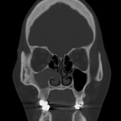
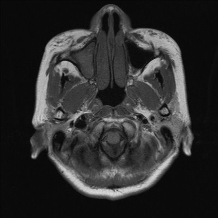
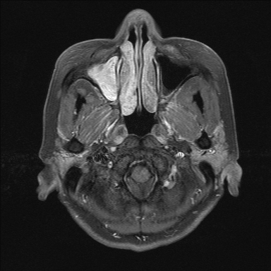
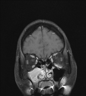
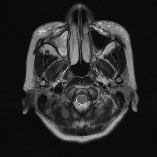
| Clinical information | 62세 여자. syncope 증세로 시행한 Brain MRI에서 우연히 발견된 right maxillary sinus의 병변. 진단은? |
| Discussion | Inverted Papilloma The term Schneiderian mucosa refers to the ectodermally derived lining of the nasal cavity and paranasal sinuses, composed generally of stratified ciliated columnar cells, loose abundant lamina propria, and minor salivary glands and their ducts. This unique mucosa may give rise to three distinct entities: fungiform or exophytic papilloma, inverted papilloma (IP), and oncocytic Schneiderian papillomas. IPs (endophytic papillomas) comprise 47% of Schneiderian papillomas and most commonly occur in males between the ages of 40 and 70 years. Characteristically, they arise from the lateral nasal wall near the middle turbinate and extend into the sinuses. This secondary extension involves the maxillary and ethmoidal sinuses, but extension into the sphenoid and frontal sinuses has been documented. Rarely, an isolated IP may arise within a sinus without any nasal involvement. Inverted papillomas rarely arise from the nasal septal wall, and fewer than 4% occur bilaterally. The most common presenting symptoms are nasal obstruction, epistaxis, and anosmia. Secondary sinusitis and tumor extension into the sinuses and orbits can cause pain, purulent nasal discharge, proptosis, diplopia, and a nasal vocal quality. Mere polypectomy results in high recurrence rates that vary from 27% to 73%. Lateral rhinotomy with en bloc resection of the lateral nasal wall and mucosa is the preferred procedure for all but the smallest localized lesions. This more extensive surgical approach has decreased recurrence rates to 0% to 14%, with most relapses occurring within 2 years. Carcinoma-ex-IP has been reported in 3% to 24% (average: 13%) of cases. Carcinoma may be concurrent with, or develop subsequent to, IP. Most reported malignancies are SCCs, but verrucous carcinoma, mucoepidermoid carcinoma, spindle cell carcinoma, clear cell carcinoma, and adenocarcinomas may also occur.10 Imaging of Schneiderian Papillomas The imaging findings for all of these papillomas can vary from a small nasal polypoid mass to an expansile nasal mass that has remodeled the nasal vault and extended into the sinuses, causing secondary obstructive sinusitis The MR and CT scan findings are nonspecific. Although apparent calcifications have been reported within the IPs, these radiodensities in reality are residual bone fragments. Nonetheless, the imaging differential diagnosis of a unilateral polypoid mass with apparent calcifications must include IPs. The nasal septum usually remains intact, but it may be bowed to the opposite side by the mass. In patients with prior surgery, including a medial antrectomy for an IP, any polypoid mass that bridges the antral–nasal border on imaging must be considered a recurrence. A unilateral mass localized to the lateral nasal wall and the middle meatus region is predictive of IP. A lobulated surface pattern, also typical, was noted in 19 of the 29 CT scans of cases. Although a septal-based solitary polypoid mass could be any of these papillomas, the location most strongly suggests a fungiform papilloma. When an area of aggressive bone destruction is seen along the margin of an IP, the radiologist must raise the possibility of an associated carcinoma. The MR imaging findings of IPs have been described as either nonspecific or highly suggestive of the lesion if there is a convoluted, cerebriform appearance on T2-weighted images and/or on T1-weighted contrast-enhanced images. Although this pattern is highly suggestive of IP, it can also be seen in other tumors such as adenocarcinoma. If necrosis is seen with a mass thought to be an IP or if there is an area of adjacent infiltrative bone invasion, coexistent carcinoma should be suggested. Reference: Head and Neck Imaging 5th ed. Peter M. Som and Huge D. Curtin. St Louis, MO: Mosby, 2011. |
| Correct answer |
Correct Answer
Semi-Correct Answer |