HOME > Weekly Case > Weekly Case
Weekly Case
종결
Case No. 359
2015-01-01~2015-01-31
출제자 : 임현경
Hit : 352
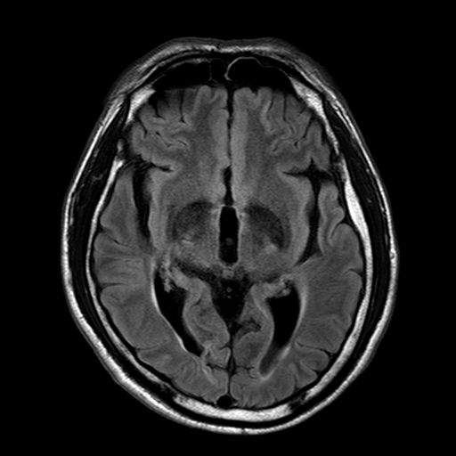
| Period | 2015-01-01~2015-01-31 |
|---|---|
| Diagnosis | Amyotrophic lateral sclerosis (Lou Gehrig’s disease) |
| Clinical information | 53세 남자환자. 5일전부터 양쪽 다리에 힘이 빠지는 증상으로 내원. 가장 적절한 진단은? |
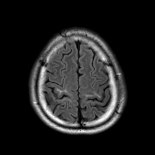
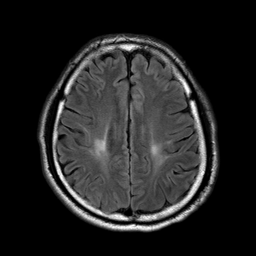
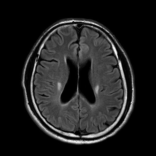
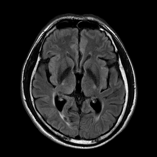
| Discussion | Review: Etiology, EPIDEMIOLOGY AND DEMOGRAPHICS ALS is characterized by progressive degeneration of motor neurons in both the brain and spinal cord. ALS has an incidence of 1-2 per 100,000 per year and is the most common motor neuron disease, representing approximately 85% of all cases. ALS is mostly sporadic; 10-15% of cases are familial. The average age of onset in familial ALS is 10 years earlier than in sporadic ALS. PRESENTATION Signs of both UMN and LMN disease are generally required for the clinical diagnosis of ALS. Disease onset is typically insidious, as at least 30% of anterior horn cells are lost before weakness becomes clinically apparent. NATURAL HISTORY Although median survival from diagnosis to death is between three and four years, 10% of patients survive beyond 10 years. Death is generally from respiratory failure due to diaphragm weakness. Imaging MR FINDINGS Macroscopic atrophy on T1WI is uncommon in ALS. Voxel-based morphometry may demonstrate subtle gray matter atrophy in the precentral gyri. Patients with ALS/FTLD exhibit a more pronounced frontotemporal volume loss. The corticospinal tract (CST) and subcortical WM appear normal in the majority of ALS patients with predominant UMN signs! A small percentage demonstrate CST hyperintensity on PD- and T2-weighted or FLAIR sequences. The hyperintensity can occur anywhere from the subcortical WM to the cerebral peduncles and pons. Changes are usually most prominent in the posterior limbs of the internal capsules and cerebral peduncles. As the CST is normally slightly hyperintense, this finding lacks both sensitivity and specificity as an imaging "biomarker" for ALS. No matter the intensity of the CST, DTI shows reduced FA in the internal capsules of ALS patients, indicating loss of microanatomical integrity. The extramotor WM also often shows decreased FA. Tractography demonstrates subcortical truncation of the CST in patients with demonstrable hyperintensity on PD- or T2-weighted sequences. MRS is generally nonspecific with decreased NAA:Cr in the precentral cortex. |
| Correct answer |
Correct Answer
Semi-Correct Answer |