HOME > Weekly Case > Weekly Case
Weekly Case
종결
Case No. 351
2014-10-01~2014-10-31
출제자 : 윤라경
Hit : 307
| Period | 2014-10-01~2014-10-31 |
|---|---|
| Diagnosis | Hematoma mimicking lymphoma |
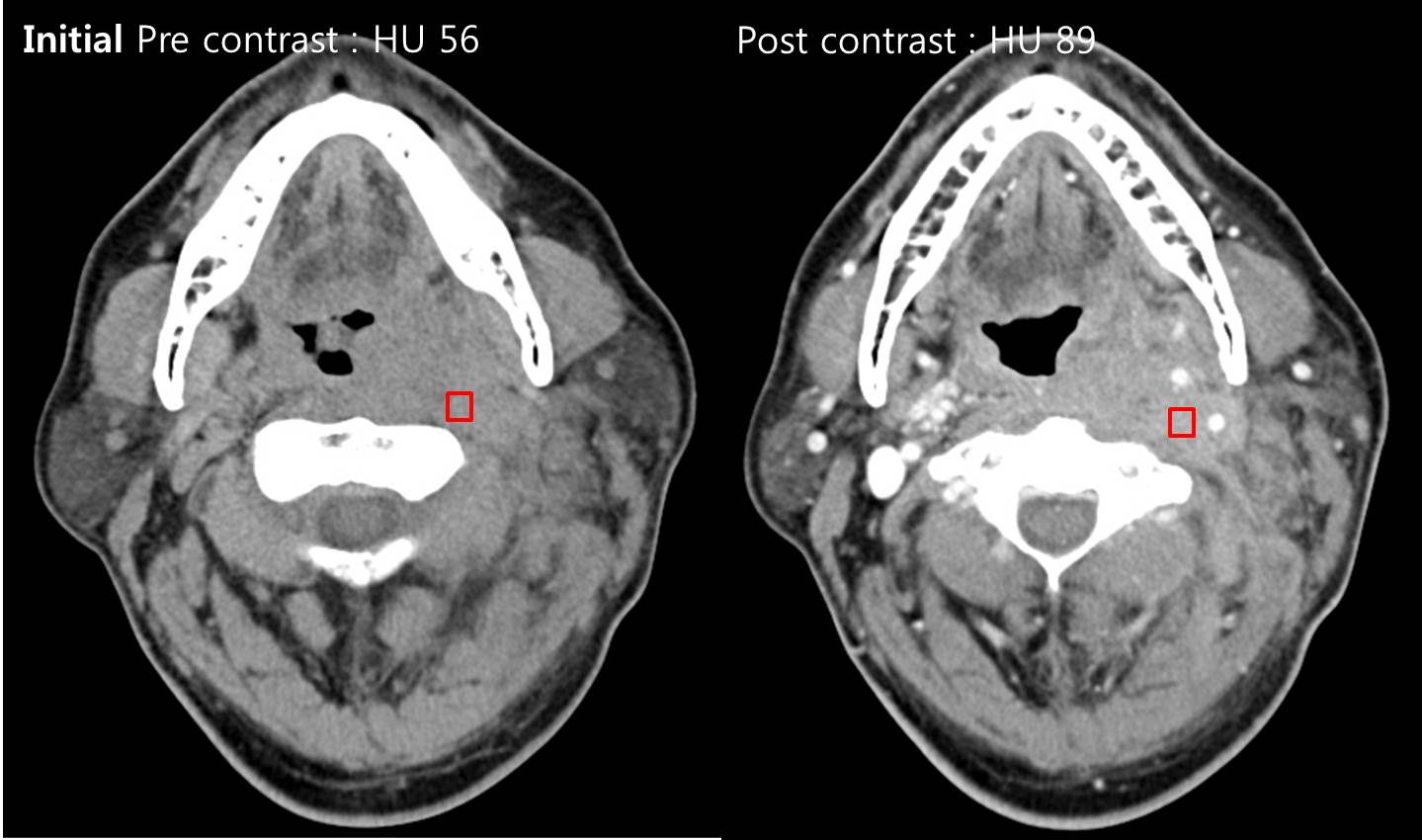
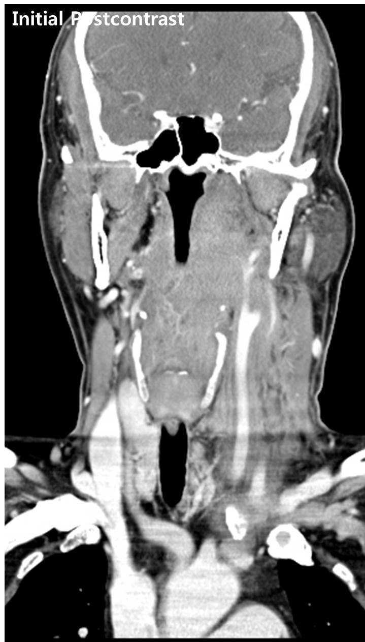
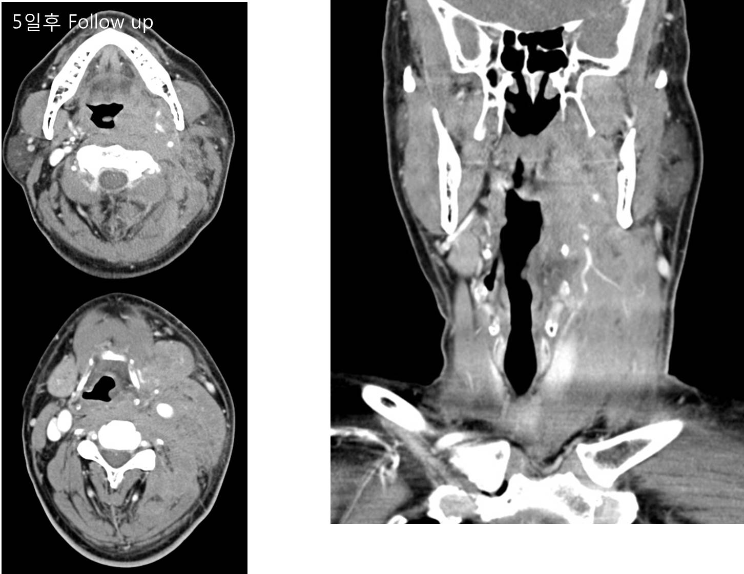
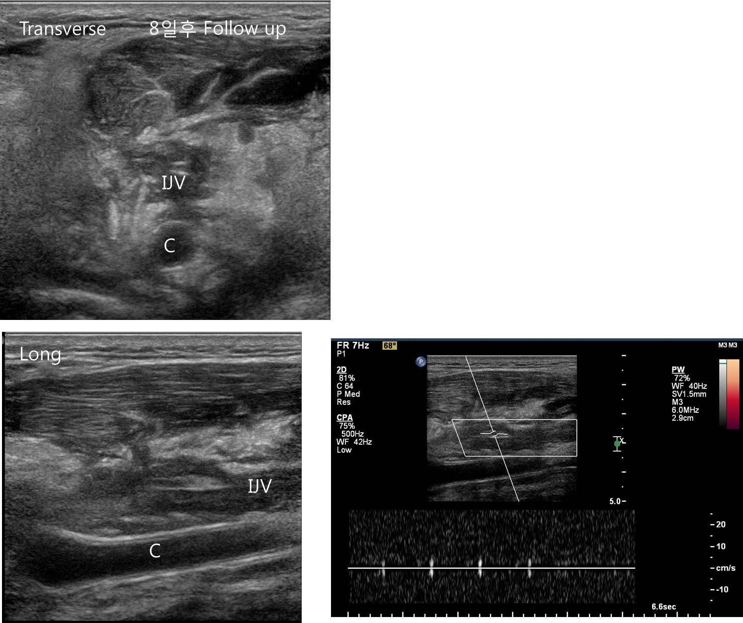
| Clinical information | M/57, neck pain으로 응급실 내원한 환자로, 내원 1주일전 febrile sense, 5일 전부터 left side neck swelling 점점 심해진 history가 있음. 진단은? |
| Discussion | ER 내원하는 환자중 원인이 head and neck neoplasm 일 수 있는데 이는 palpable mass, swelling, compromised airway 때문일 수 있음. 대부분은 squamous cell CA이나 lymphoma도 상당 부분 차지함. Most common site of lymphoma involvement is Waldeyer ring (i.e., lingual, palatine, and nasopharyngeal tonsils) in head and neck. Imaging Findings of Lymphoma CT -high denstiy infiltrative lesion at the left carotid space with extension to subamandibular, anterior and posterior cevical spaces, parapharyngeal and retropharyngeal spaces - increased extent on follow up image - no definite evidence of extravasation of contrast (clue) - encasement of the left jugular vein US - Diffuse hyperechoic infiltrative lesion mainly involving the left carotid space and the adjacent soft tissue and neck spaces - Diffuse hypoechoic lesion encircling the collapsed left internal jugular vein and its tributary MR - Isodense to muscle, normally homogeneous with moderate contrast enhancement - MR- T1WI hypo or isointense to muscle, variable enhancement Reference) Adam G et al. Extranodal lymphoma from head to toe radio clin North Am 2008 Mar;46(2):363-78 Benjamin Ludwig, et al. Diagnostic Imaging in Nontraumatic Pediatric Head and Neck Emergencies Radiographics 2010, vol.30: 781-799 |
| Correct answer |
Correct AnswerSemi-Correct Answer |