HOME > Weekly Case > Weekly Case
Weekly Case
종결
Case No. 350
2014-10-01~2014-10-31
출제자 : 윤라경
Hit : 372
| Period | 2014-10-01~2014-10-31 |
|---|---|
| Diagnosis | Neuro-Behcet disease |
| Clinical information | 33/M C.C: Sudden onset diplopia Rt. Side hypesthesia |
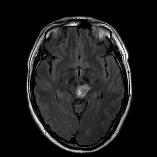
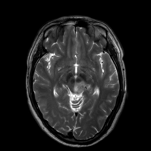
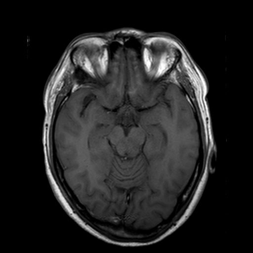
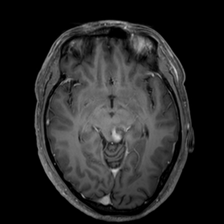
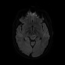
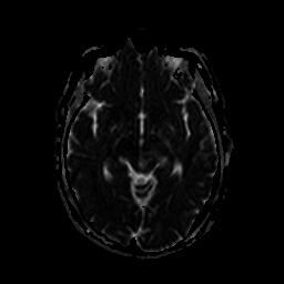
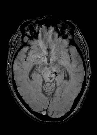
| Discussion | MR 판독: About 2cm sized, T2 high, T1 low SI lesion with perilesional edema in left midbrain. Dark SI and blooming artifact on susceptibility weighted image. Partial rim like enhancment. No direct communication with cerebral vessels on MR. --> r/o chronic hemorrhage d/t venous infarction or small AVM > cavernous hemangioma, more likely. r/o vasculitis. Disease review: Brainstem involvement (50%) > thalamus, basal ganglia > cerebral hemispheric white matter Small circular, linear, crescent-shaped, or irregular foci of T2/FLAIR hyperintensity in the midbrain Mass effect is usually minimal except acute phase SWI : hemorrhagic foci Commonly patchy enhancement Rarely, strong, uniform enhancement References> 1) S. Albaryram, S. Saip, Z.I. Hasiloglu, Et al. Evaluation of parenchymal Neuro-Behçet disease by using susceptibility-weighted imaging. AJNR. 2001;32:1050-1055 2) Kocer N, Islak C, Siva A, et al. CNS involvement in Behçet’s disease syndrome: an MR study. AJNR 1999;20:1015-24 3) Anne G. Osborn. Osborn’s Brain: Imaging, Pathology, And Anatomy. 276~277 |
| Correct answer |
Correct Answer
Semi-Correct Answer |