HOME > Weekly Case > Weekly Case
Weekly Case
종결
Case No. 349
2014-09-01~2014-09-30
출제자 : 이영흔
Hit : 326
| Period | 2014-09-01~2014-09-30 |
|---|---|
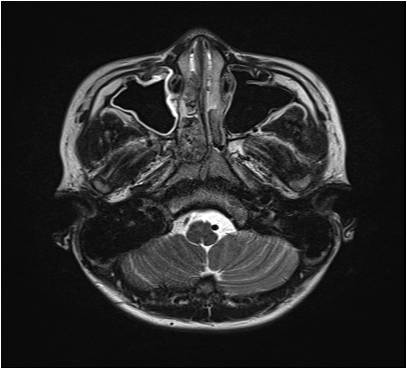
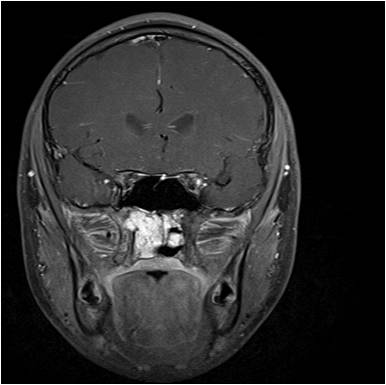
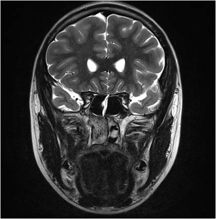
| Diagnosis | Angiofibroma |
| Clinical information | 16/M Nasal obstruction (O : 2MA) |
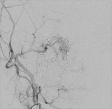
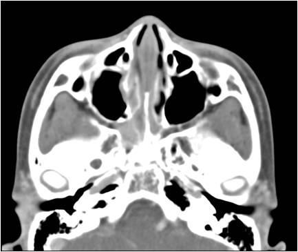
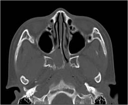
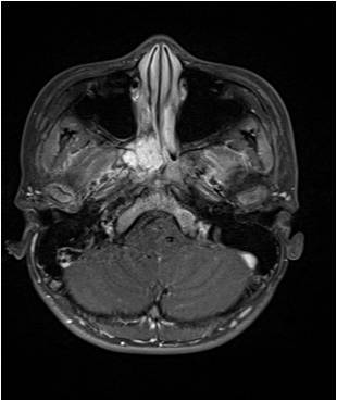
| Discussion | 수술적 진단명: angiofibroma **exclusive하게 2-25세의 청소년 남자에서 생기며, Nasal obstruction(90%), epistaxis(60%)의 증상을 보인다. Androgen, dihydrotestosterone 등 남성호르몬과 연관되어 있는 것으로 생각되어지지만, 아직 명확한 Etiology는 밝혀지지 않았다. 대개 internal maxillary artery를 main feeding artery로 하는 vascular tumor의 형태로 생기게 되고 Nasal cavity 에서 생긴 이후에 tumor가 extension 하는데 PPF (Pterygopalatine fossa)가 중요한 역할을 하여 여기에서 다양한 tract을 통해 tumor가 sinus skull base 등으로 extension을 하게 된다. tumor growth와 extension을 이해하기 위해서는 PPF의 anatomy를 이해하는 것이 매우 중요하며, PPF는 앞쪽으로는 Maxilla, 뒤쪽/roof로는 sphenoid bone으로 둘러싸여 있고 floor는 palatine으로 이루어져 있는 역피라미드 형태를 가지고 있고 angiofibroma는 PPF 의 SPF(sphenopalatine foramen)에서 origin 하거나 nasal cavity에서 들어오게 되며(NC와 PPF의 communicate) 이러한 Communication 들이 tumor가 extension는 통로 역할을 하게 되며 SPF를 통하여 nasal cavity로, pterygomaxillary fissure를 통하여 Infratempora fossa로, Foramen rotundum을 통하여 middle cranial fossa로 들어가게 되거나 IOF를 통하여 orbit으로 extension을 할 수 있다. CT에서는 SPF margin에서 origin해, 주변 bone에 remodeling이나 destruction을 시키는 enhance 잘(avid) 되는 diffuse soft tissue mass로 보이며, 종종 maxillary sinus의 posterior wall을 앞쪽으로 bowing시킬 수 있다. MR에서는 enlarged된 vessel들이 signal void의 형태로 salt and pepper의 모양을 보일수 있다. Angiography에서는 ECA(주로는 internal maxillary artery, ascending pharyngeal artery와 같은)로부터의 feeding vessel을 볼 수 있으며 capillary blush로 관찰된다. Perioperative bleeding을 줄이기 위해 술 전 embolization을 하기도 하며, sphenoidal or intracranial extension을 확인 한 후, complete surgical resection을 목표로 한다. reference: Diagnostic imaging H&N, juvenile angiofibroma II 2 64-67 |
| Correct answer |
Correct Answer
Semi-Correct Answer |