HOME > Weekly Case > Weekly Case
Weekly Case
종결
Case No. 346
2014-08-01~2014-08-31
출제자 : 차지훈
Hit : 359
| Period | 2014-08-01~2014-08-31 |
|---|---|
| Diagnosis | Hypereosinophilic syndrome |
| Clinical information | M/54, dizziness 진단은? |
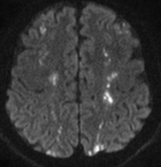
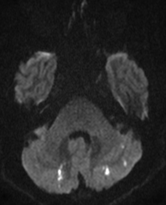
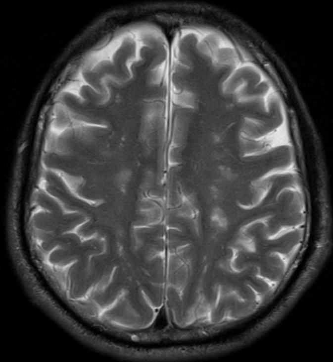
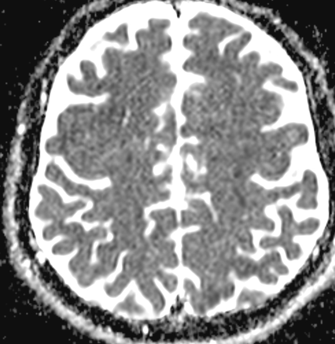
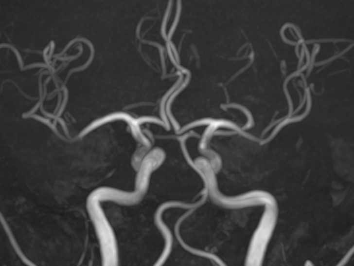
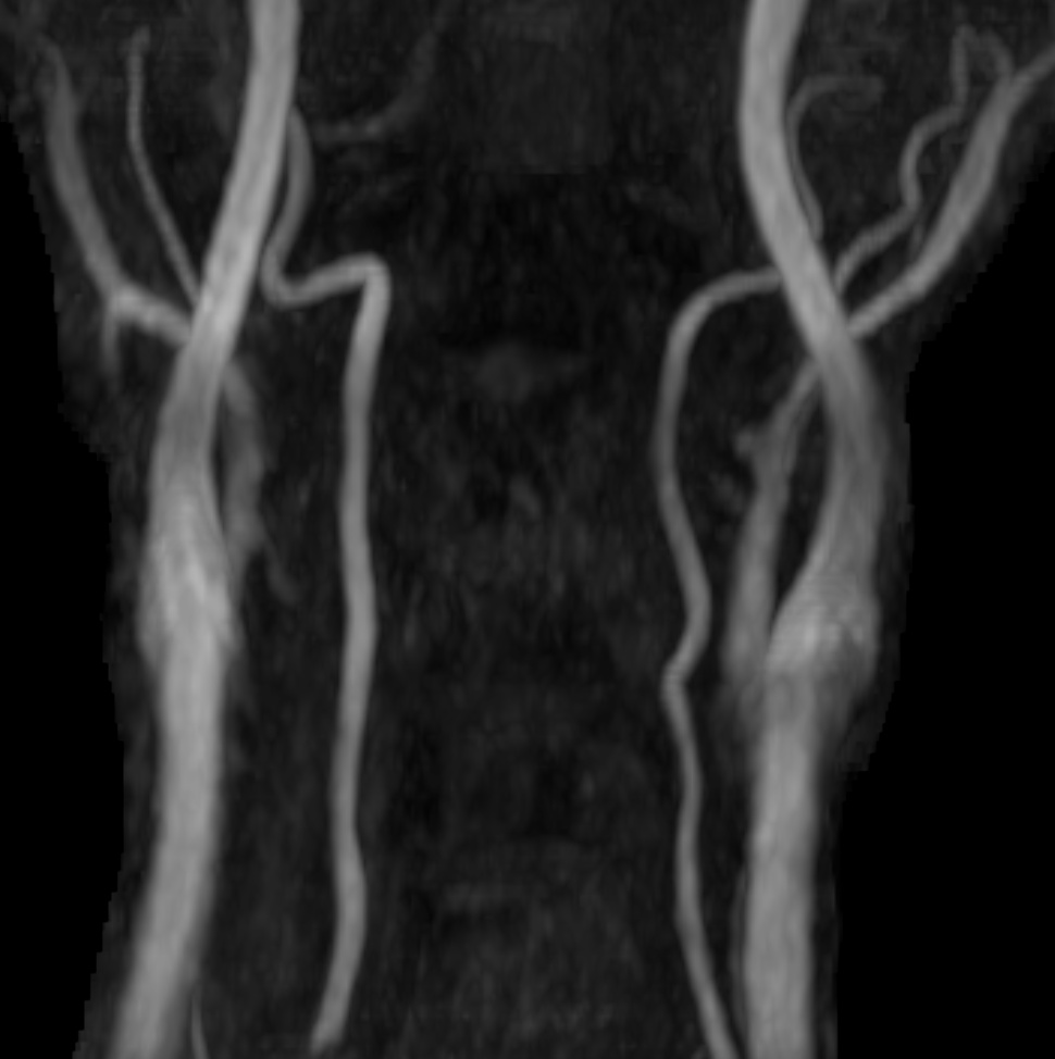
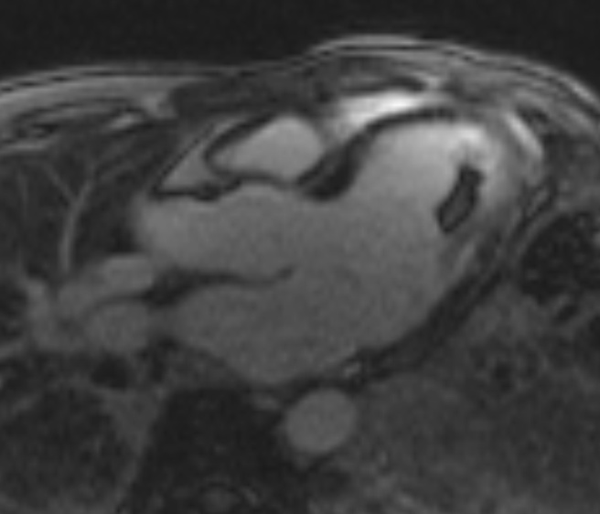
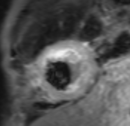
| Discussion | Imaging findings - Bilateral symmetric multistage small borderzone infarctions involving cerebrum and cerebellum - No stenoocclusive lesion of intracranial artery and proximal ICA on MRA - Thrombus in left ventricle with subendocardial enhancement - Increased eosinophil count >3000 /uL (not shown) Review - Hypereosinophilic syndrome is difined as persistent and marked blood eosinophilia (>1,500/μL, >6 months), the absence of a primary cause of eosinophilia (such as parasitic, allergic, malignant disorder, “idiopathic” for unknown pathogenesis), and evidence of eosinophil-mediated end organ damage (CNS, PNS, heart, lung, skin) - Cerebral infarction: thromboembolism and vascular endothelial toxicity of eosinophils. Microthromboemboli, border zone infarction Activated eosinophils injure human endothelial cells by releasing a number of cytopathic substances such as major basic protein Major basic protein is a potent stimulator of platelet activation and aggregation - Cardiac involvement Loeffler’s endocarditis, endomyocardial fibrosis Three stage (early necrotic stage, thrombotic stage, fibrotic stage) |
| Correct answer |
Correct Answer
Semi-Correct Answer |