HOME > Weekly Case > Weekly Case
Weekly Case
종결
Case No. 345
2014-08-01~2014-08-31
출제자 : 차지훈
Hit : 330
| Period | 2014-08-01~2014-08-31 |
|---|---|
| Diagnosis | Pilomyxoid astrocytoma |
| Clinical information | M/21, diplopia 진단은? |
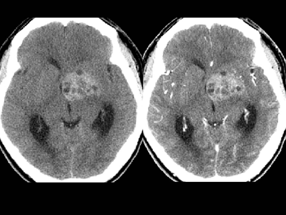
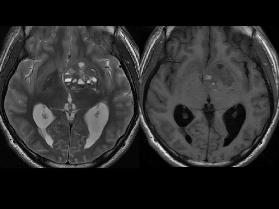
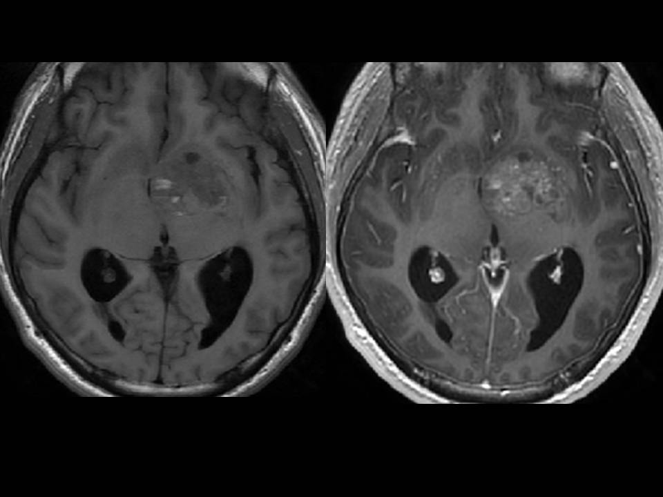
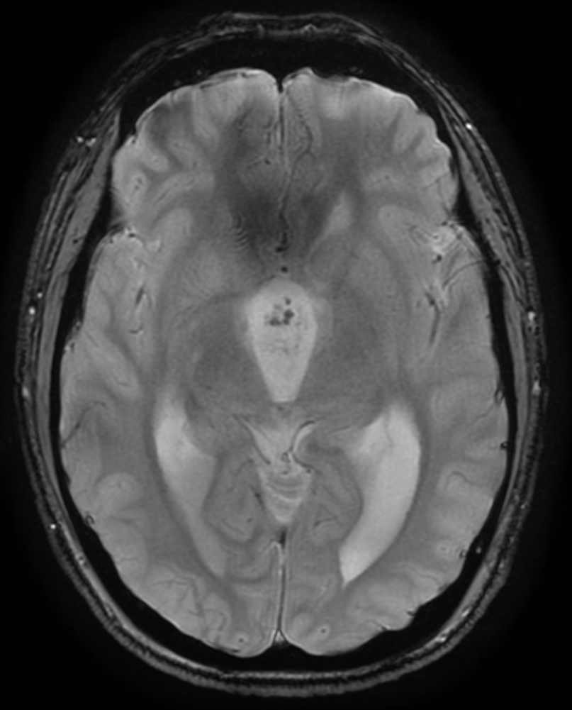
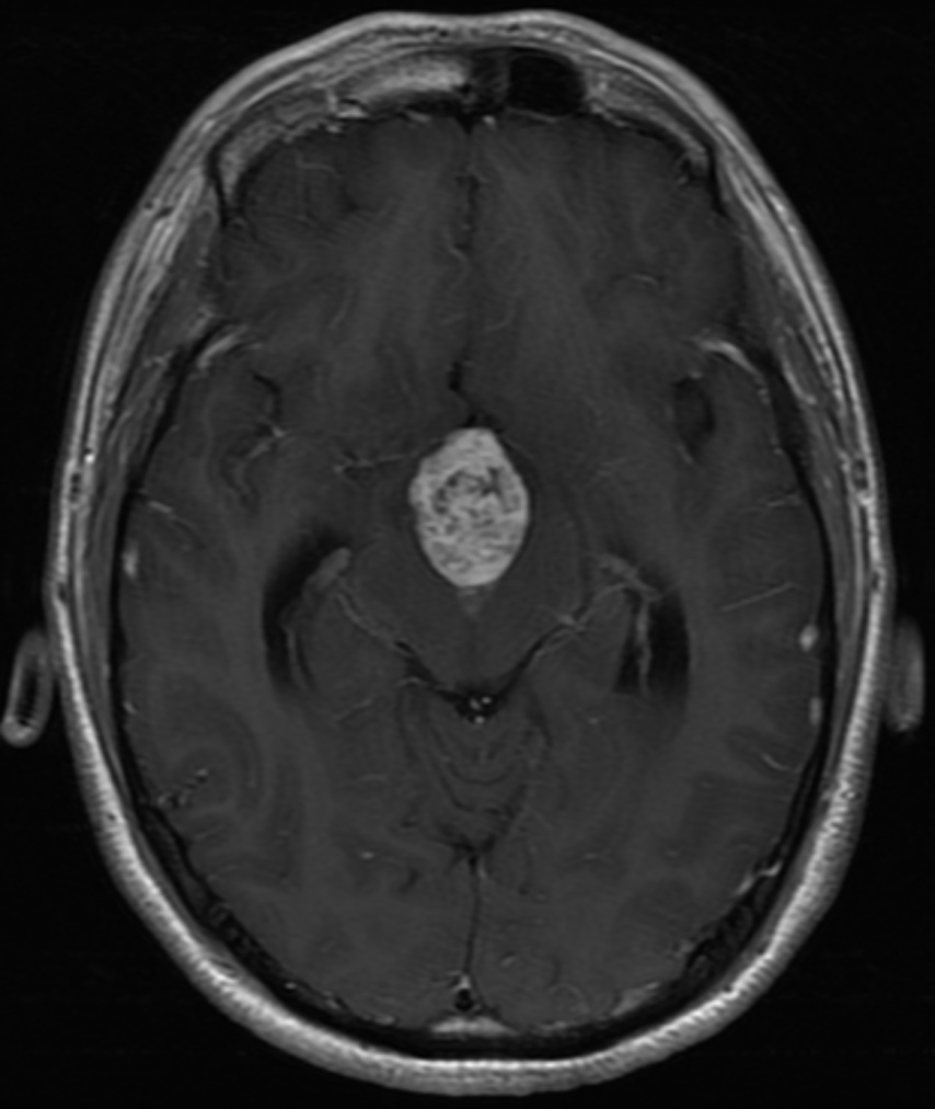
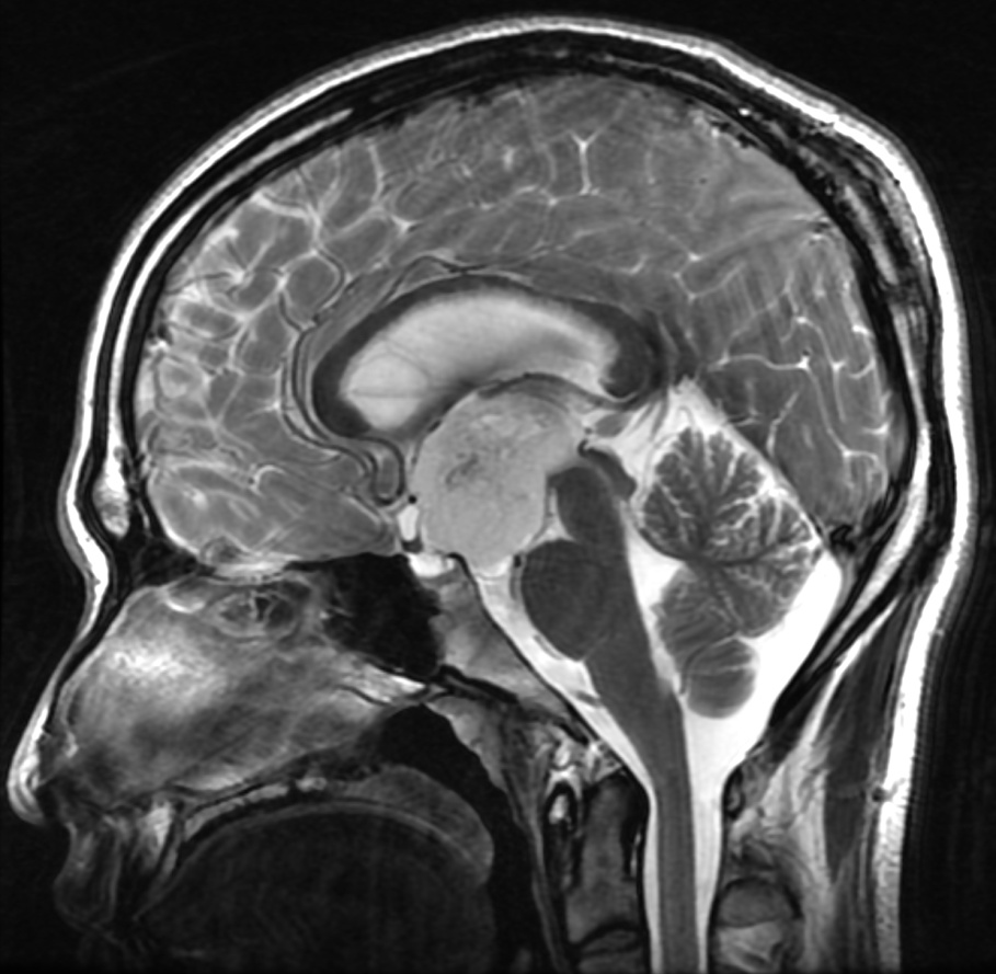
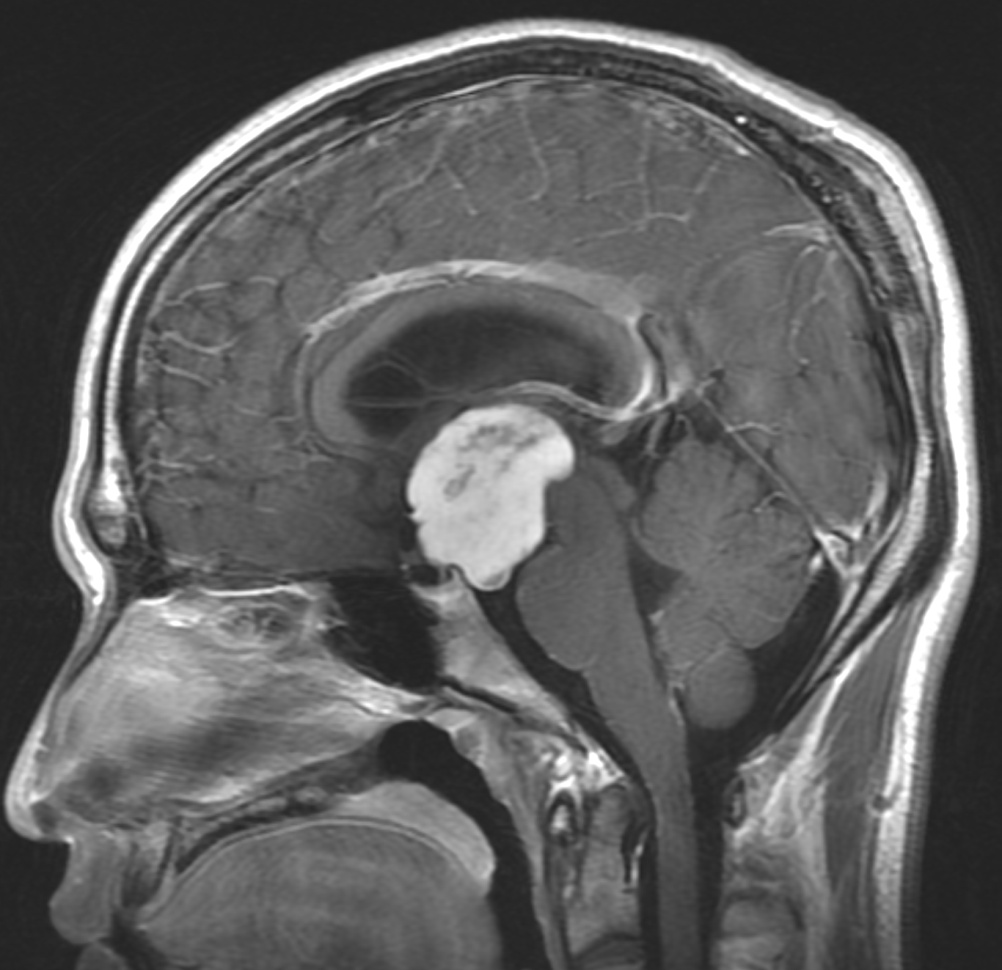
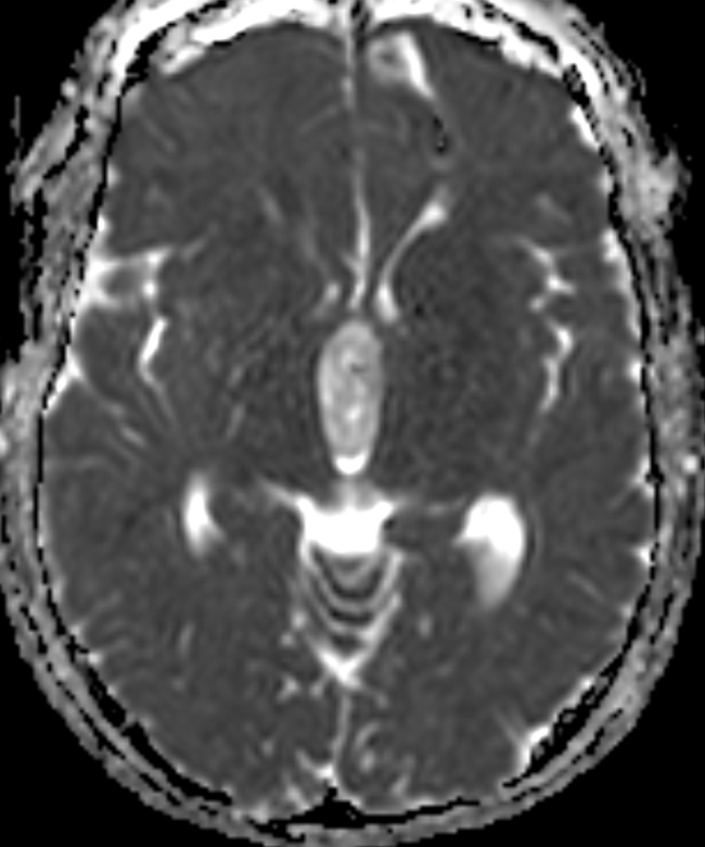
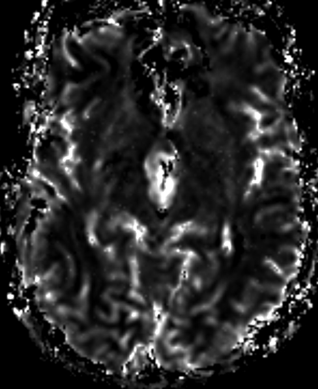
| Discussion | Imaging findings - T2 hyperintense mass involving suprasellar area and hypothalamus, protruding into third ventricle, causing obstructive hydrocephalus - Internal hemorrhage on GRE - Delayed fill in enhancement pattern suggestive of myxoid compoment - High ADC value suggestive of low cellularity, increased CBV Review - WHO grade II Occur early age, worse prognosis than pilocytic astrocytoma (grade I) Aggressive, leptomeningeal seeding can occur - Suprasellar/hypothalamic/chiasmatic/third ventricular region most common (60%) - Monomorphous piloid cell with myxoid backbround - Hyperintense T2/FLAIR, solid with central necrosis - Intratumoral hemorrhage common (10~25%), calcification uncommon (<10%) |
| Correct answer |
Correct AnswerSemi-Correct Answer |