HOME > Weekly Case > Weekly Case
Weekly Case
종결
Case No. 339
2014-06-01~2014-06-30
출제자 : 최승홍
Hit : 354
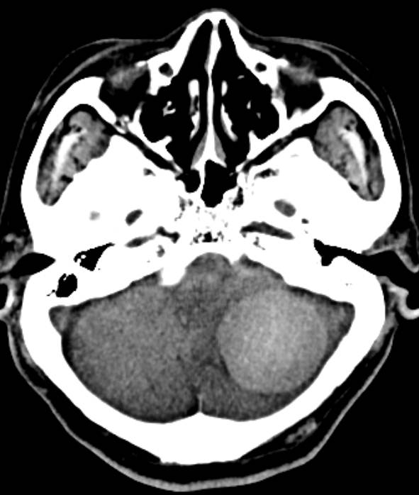
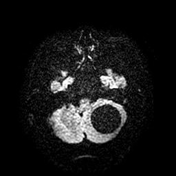
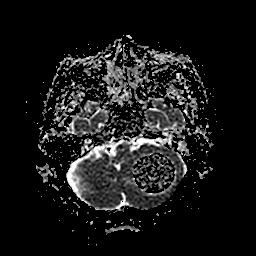
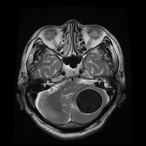
| Period | 2014-06-01~2014-06-30 |
|---|---|
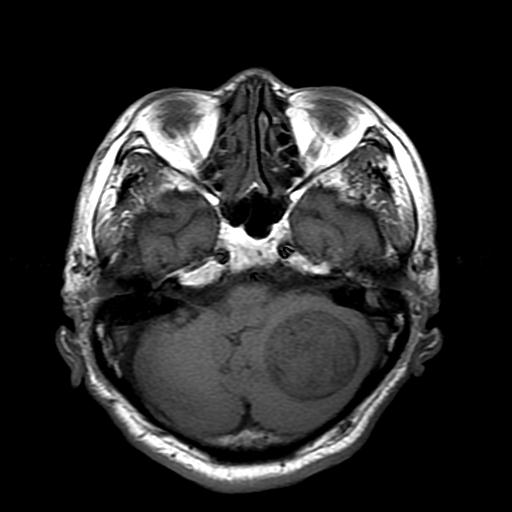
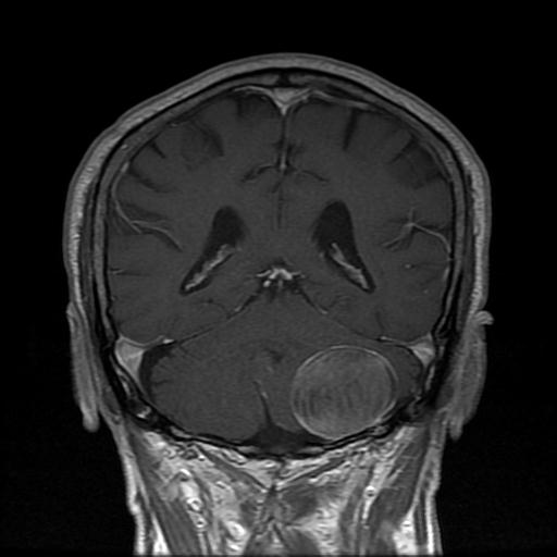
| Diagnosis | Sclerosing Meningioma |
| Clinical information | M/76 어지러움, 두 달 전부터 진단은? |
| Discussion | Background - a distinct subtype of benign meningioma that may lack the classic appearance and manifestation of typical meningothelial meningiomas. Pathology - whorl formation around sclerotic vessels, with tumor cells demonstrating glial fibrillary acidic protein expression cf. typical fibrous meningioma shows parallel and interlacing bundles of spindlelike cells embedded in a matrix of collagen and reticulin Radiologic findings - CT : poor or partial enhancement is seen and calcification is more common cf. classic meningothelial meningioma : a relatively hyperattenuating, broad based extraaxial mass with a smooth or lobulated outline that enhances homogeneously with contrast material administration - MR : T2-weighted MR imaging demonstrates a mass with low signal intensity, which corresponds to the psammomatous calcification seen at histopathologic analysis Reference> RadioGraphics 2003; 23:785–789 |
| Correct answer |
Correct AnswerSemi-Correct Answer |