HOME > Weekly Case > Weekly Case
Weekly Case
종결
Case No. 326
2014-01-01~2014-01-31
출제자 : 최진욱
Hit : 375
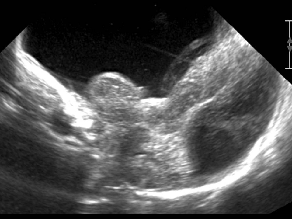
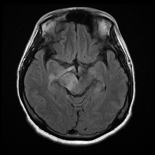
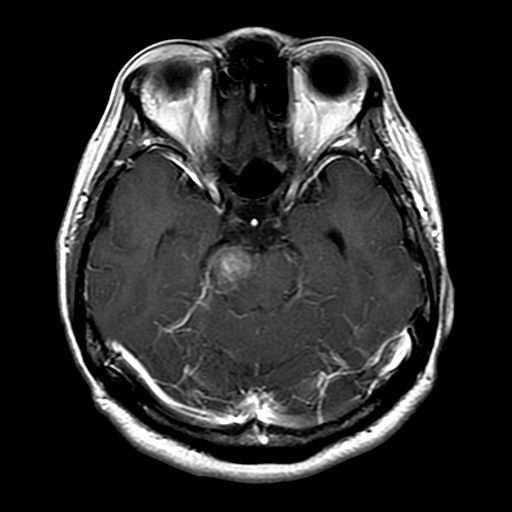
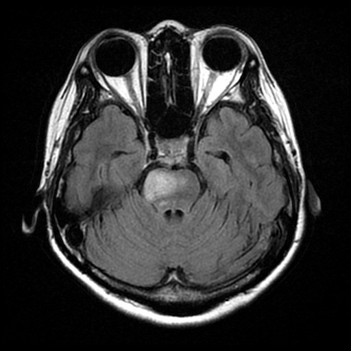
| Period | 2014-01-01~2014-01-31 |
|---|---|
| Diagnosis | Neuro behçet disease |
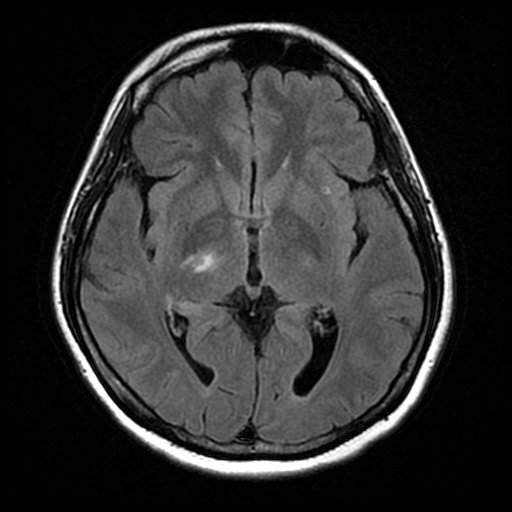
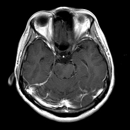
| Clinical information | F/45 C.C : headache(VAS 9), progressive after several days, Lt. side weakness, dysmetria, anisocoric pupil, Lt. side increased DTR 아래 3개의 사진 : 일주일 후 follow-up MRI |
| Discussion | Neuro behçet disease Chronic, idiopathic, relapsing-remitting multisystemic vascualr-inflammatory disease characterized by recurrent orogenital uncerations and uveitis CNS inbolved in up to 20% of pts Imaging of Neuro behçet disease T2 high SI on Brainstem: ventral midbrain(cerebral peduncles) and pons typically invovled Basal ganglia>thalami>WM(subcortical) Sinus thrombosis, aterial occlusion, aneurysm DWI : variable, restriction in acute phase T1C+ : patch enhacement Reversible course Chronic brainstem atrophy wtihout cerebral atrophy ddx from the other vasculitis such as SLE |
| Correct answer |
Correct Answer
Semi-Correct Answer |