HOME > Weekly Case > Weekly Case
Weekly Case
종결
Case No. 324
2013-12-02~2013-12-31
출제자 : 안성수
Hit : 356
| Period | 2013-12-02~2013-12-31 |
|---|---|
| Diagnosis | Giant cell tumor of tendon sheath at TMJ (PVNS) |
| Clinical information | 73/F CC: Tinnitus 진단은? |
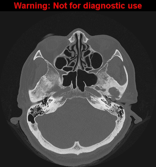
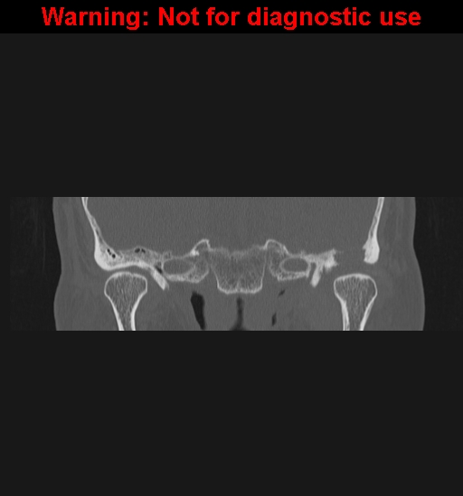
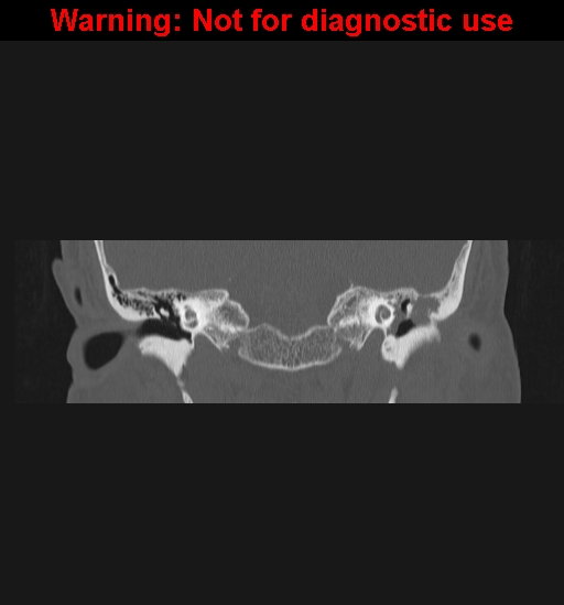
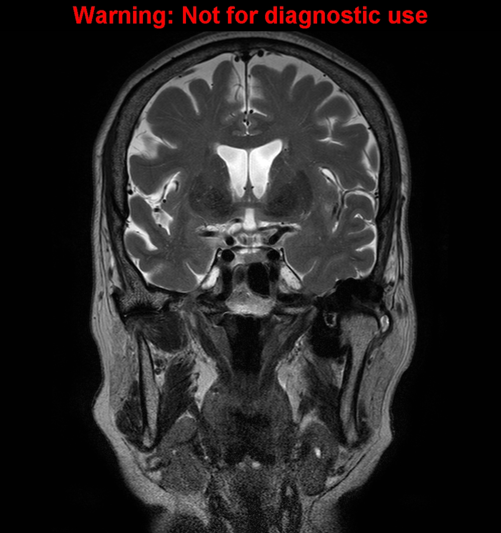
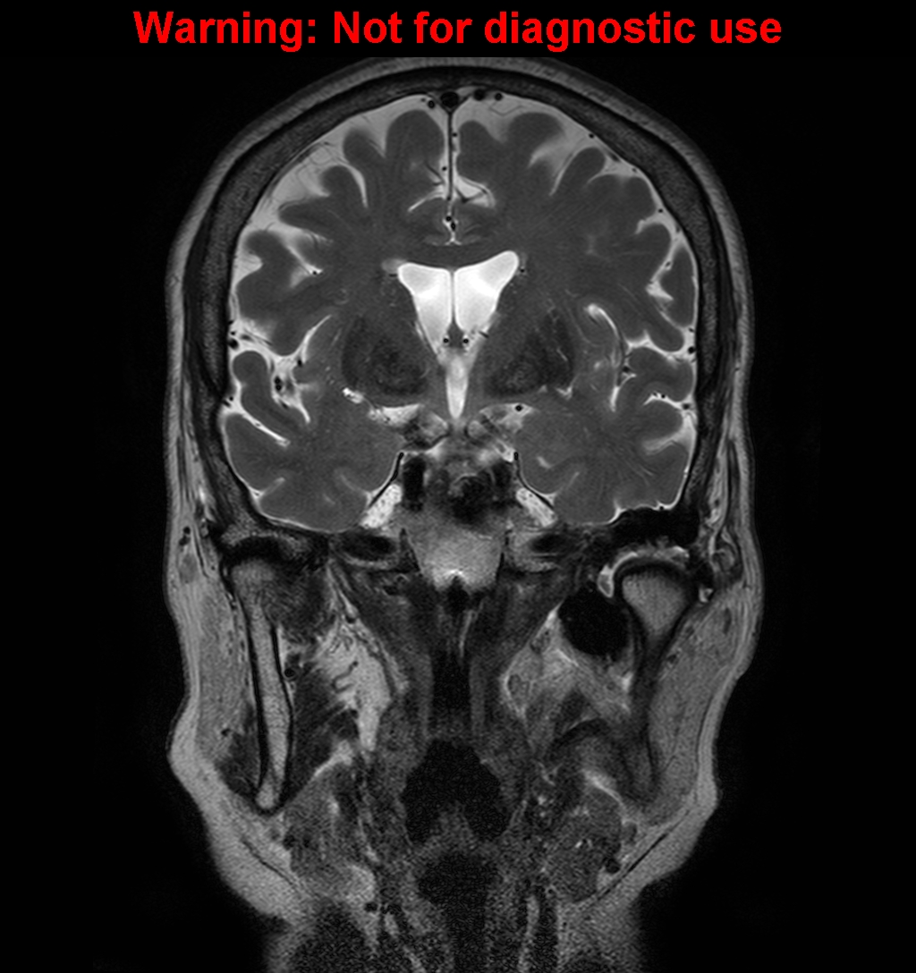
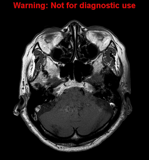
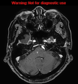
| Discussion | 정답: Giant cell tumor of tendon sheath at TMJ (PVNS) 해설: Temporal bone CT coronal images show lobulated erosion of glenoid fossa of TMJ extending into the anterior EAC. T1WI and T2WI demonstrate marked hypointense lesion in the glenoid fossa and small amount of effusion in the left TMJ. The lesion show mild enhancement. Benign but locally aggressive, proliferative disorder of synovium, also known as pigmented villonodular synovitis CT: lobulated or round erosion of mandibular condyle with/without glenoid fossa MRI: hypointense to isointense on T1WI, hypointense due to hemosiderin deposition on T2WI, mild enhancement |
| Correct answer |
Correct Answer
Semi-Correct Answer |