HOME > Weekly Case > Weekly Case
Weekly Case
종결
Case No. 323
2013-12-02~2013-12-31
출제자 : 안성수
Hit : 328
| Period | 2013-12-02~2013-12-31 |
|---|---|
| Diagnosis | chondrosarcoma |
| Clinical information | 32/M CC: headache, syncope 진단은? |
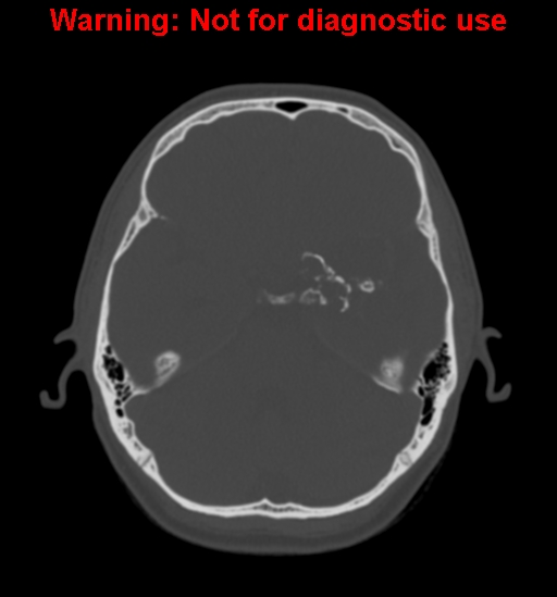
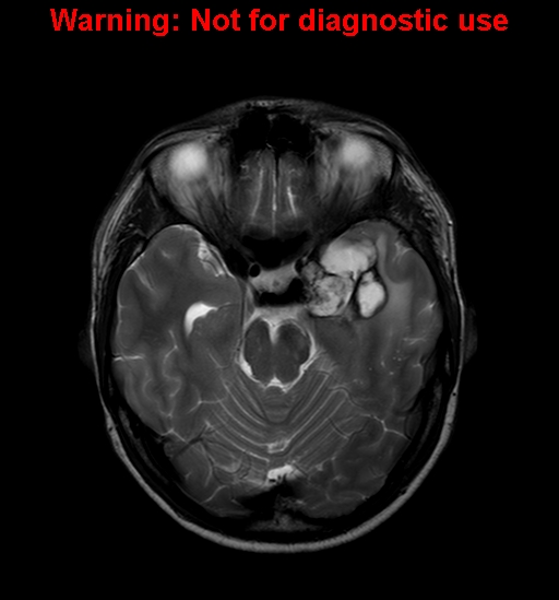
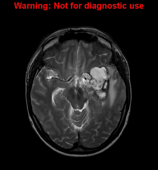
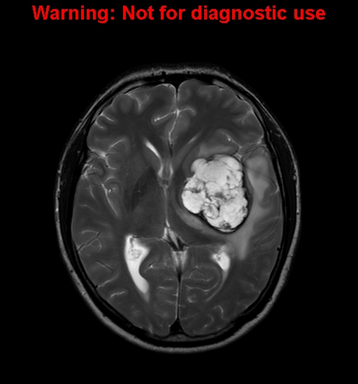
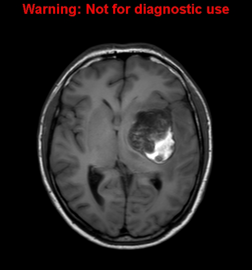
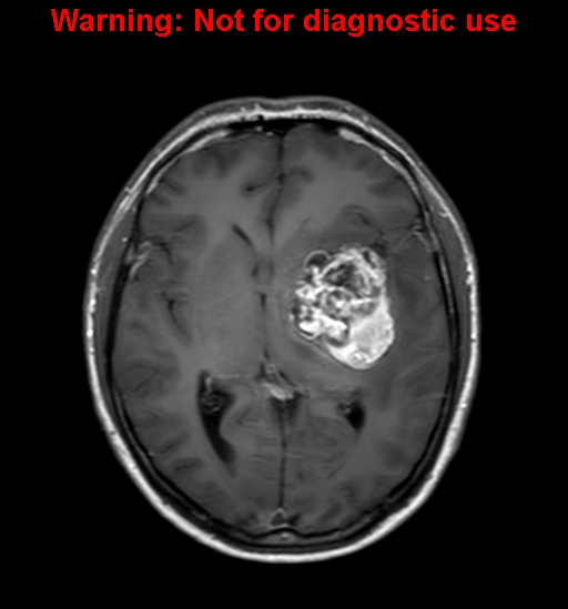
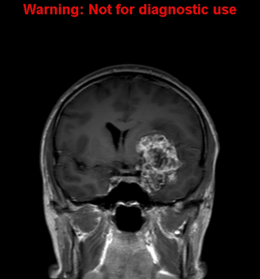
| Discussion | 정답 : chondrosarcoma 해설: T2WI demonstrates hyperintense mass with hypointense rim and septa. The mass shows extraaxial location and is probably arising from the central skull base. Subacute hemorrhage is seen in the posterior part of the mass on T1WI and the mass enhances heterogeneously. Noncontrast CT demonstrates ring and arc calcifications in the Lt paraclinoid region. Location: petro-occipital fissure (2/3), anterior basisphenoid (1/3) Bone CT: characteristic ring and arc calcifications in tumor matrix (50%) MRI: low to intermediate SI on T1WI, high SI with scattered hypointense foci due to calcifications on T2WI, heterogeneous enhancement |
| Correct answer |
Correct Answer
Semi-Correct Answer |