HOME > Weekly Case > Weekly Case
Weekly Case
종결
Case No. 313
2013-08-01~2013-08-31
출제자 : 황승배
Hit : 524
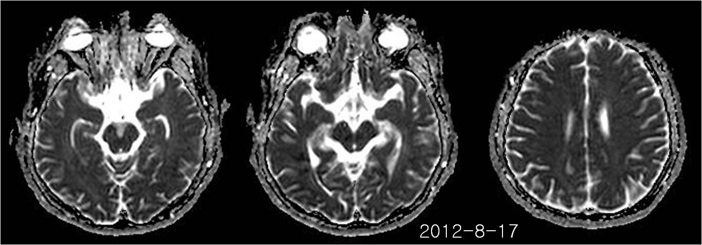
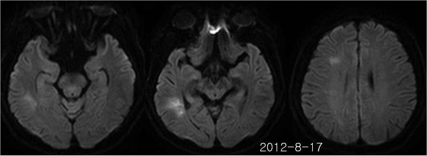
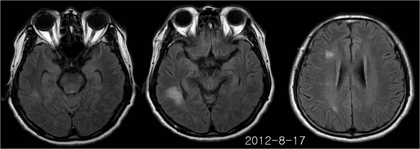
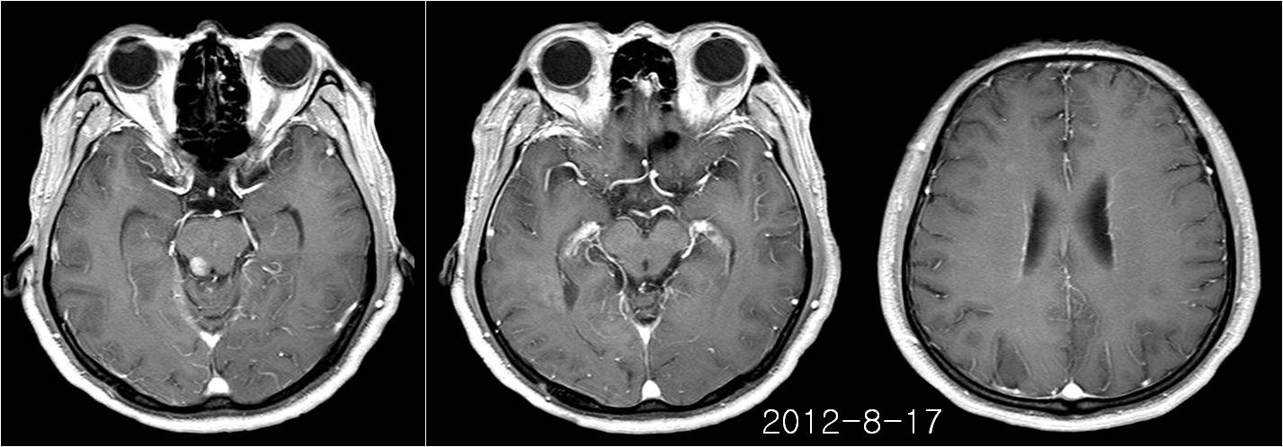
| Period | 2013-08-01~2013-08-31 |
|---|---|
| Diagnosis | Intravascular large B cell lymphomatosis |
| Clinical information | 63/M C/C : Dysarthria, apraxia 진단은? |
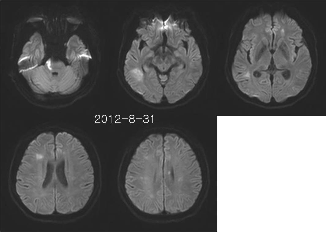
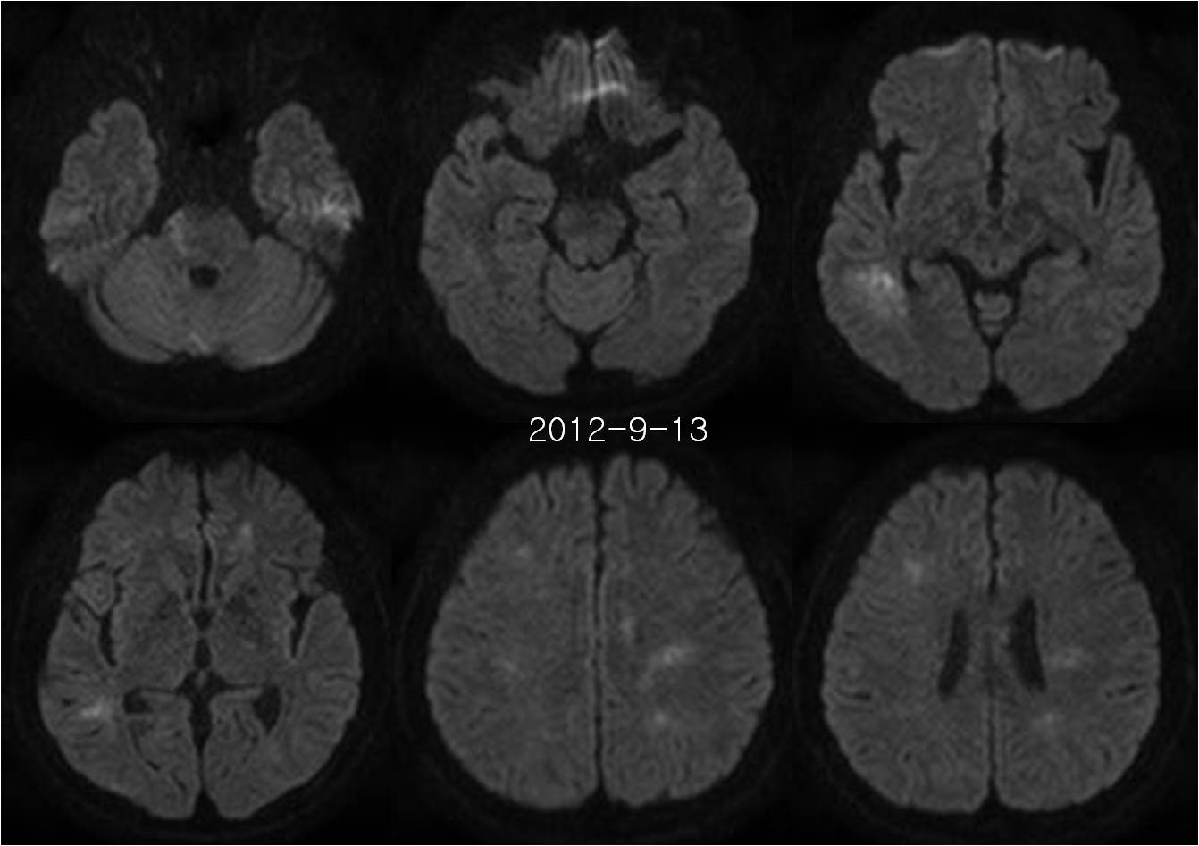
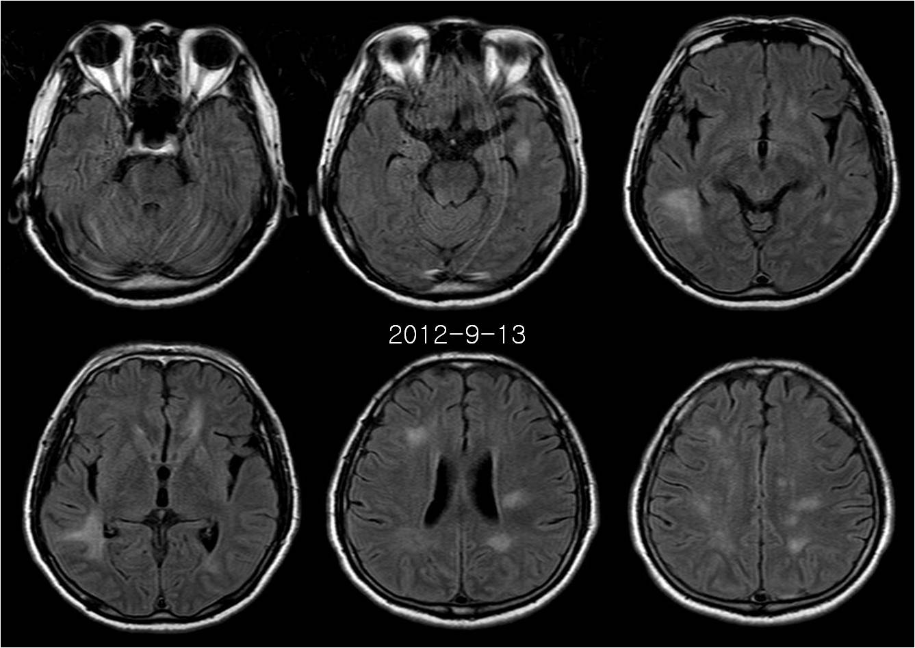
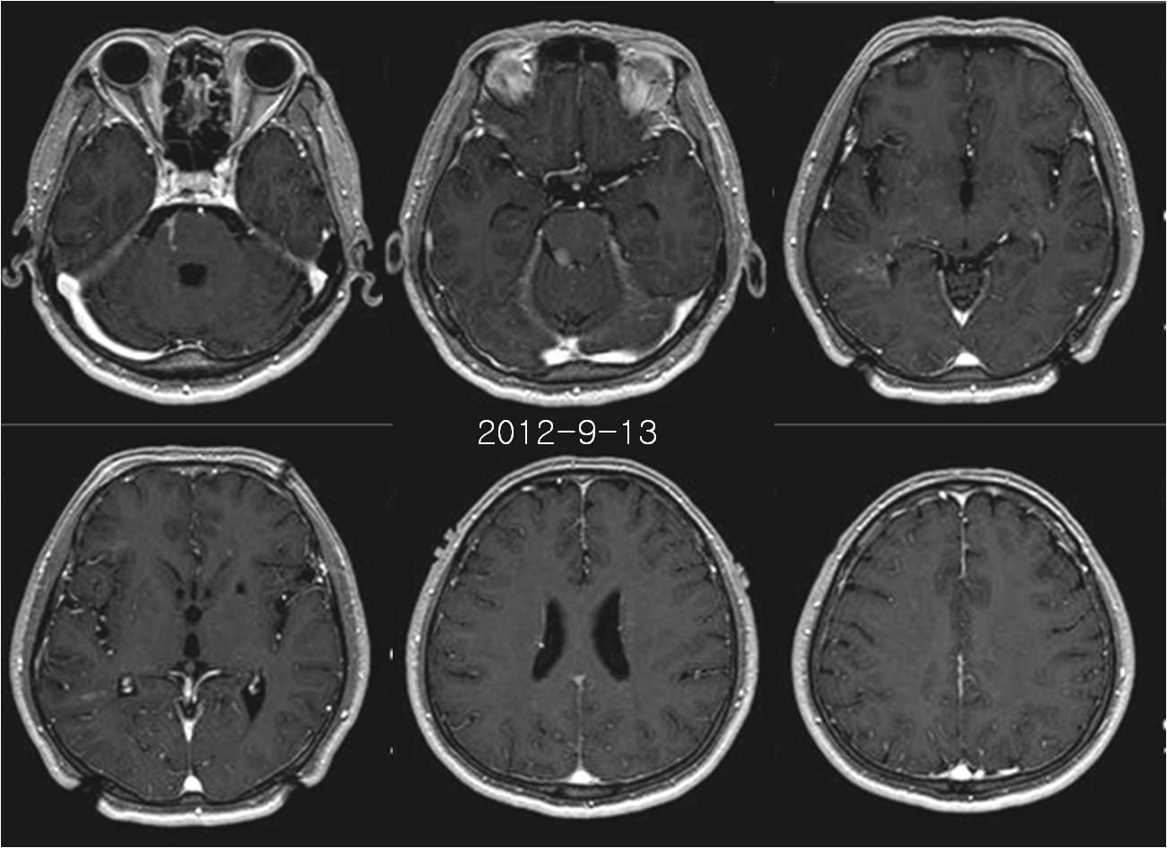
| Discussion | Intravascular large B cell lymphomatosis 63/M C/C : Dysarthria, apraxia 영상소견 - 초기 영상 (2012-8-17) 에서 FLAIR상 Rt temporal lobe, Rt frontal lobe, Rt parietal lobe에 multifocal high signal lesions이 관찰되며 병변은 white matter를 침범하고 있음. DWI에서 Rt temporal lobe lesion은 diffusion restriction을 보이나 다른 병변들은 명확한 diffusion restriction소견 보이지 않음. 조영증강영상에서 병변들은 subtle or minimal enhancement가 의심됨. Rt dorsal pons에 FLAIR & DWI에서 잘 보이지 않았던 well-defined strong enhancing nodular lesion이 있음. - 14일 후 DWI MR (2012-8-31)에서 이전 병변들은 큰 변화없이 보이고 있으나 Rt lateral pons, body of corpus callosum, Lt frontal lobe white matter, and Lt periventricular white matter에 새로운 병변들이 관찰됨. - 27일 후 MR (2012-9-13) 영상에서 이전 병변들 외에 Lt temporal lobe, Lt periventricular white matter, and both centrum semiovale부위에 새로운 multifocal lesion들이 생겼으며 diffusion high signal을 보이고 있음. - Stereotatic biopsy를 통해 intravascular large B cell lymphomatosis로 확진되었음. Intravascular large B cell lymphomatosis Rare systemic disease with a predilection for the CNS and skin Form of large-cell non-Hodgkin lymphoma that is characterized by angiotropic growth Pathologic diagnosis of IVL may be difficult and requires immunologic analysis and electron microscopy Clinical presentation & Diagnostic imaging - nonspecific Diagnosis should be considered in patients presenting with nonfocal neurologic deficits or rapidly progressive dementia without documented cerebrovascular disease or immunosuppression Advanced stage at presentation - overall mortality rate > 80% - average survival time of only 9-13 months Recognition of the MR findings may lead to prompt biopsy, allowing earlier diagnosis Angiography and CT - multiple vascular occlusions and stroke MR findings - arise in part from vessels occluded by intravascular lymphomatosis - producing a wide spectrum of abnormalities that are characteristically multifocal Characteristic MR imaging findings • 1) T2 HSI on in the deep white matter ( 45%) - single most common imaging finding - nonspecific nature - d/t small-vessel occlusion leading to ischemia 2) Infarct-like lesions (36%) - pathologic finding of multiple areas of recent or resolving infarction 3) Enhancing parenchymal mass lesions - mimicking other forms of lymphoma - focal nodular enhancement in callosum ; represent extravascular spread of tumor. - extensive meningeal enhancement ; direct infiltration of meningeal blood vessels ; microinfarcts of the meninges Reference William RL, Meltzer CC, Smirniotopoulos JG, Fukui MB, Inman M. Cerebral MR imaging in intravascular lymphomatosis. AJNR Am J Neuroradiol 1998:19;427-431 |
| Correct answer |
Correct Answer
Semi-Correct Answer |