HOME > Weekly Case > Weekly Case
Weekly Case
종결
Case No. 310
2013-07-01~2013-07-31
출제자 : 이영흔
Hit : 419
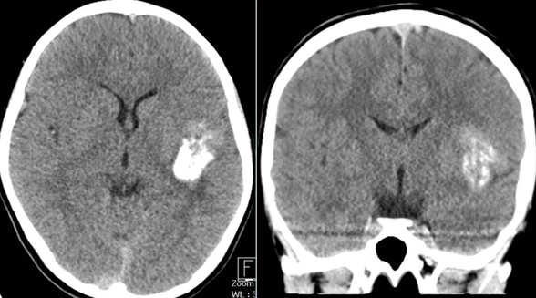
| Period | 2013-07-01~2013-07-31 |
|---|---|
| Diagnosis | Meningioangiomatosis |
| Clinical information | M/10, GTC like seizure |
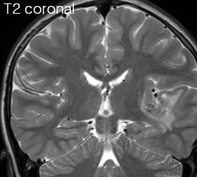
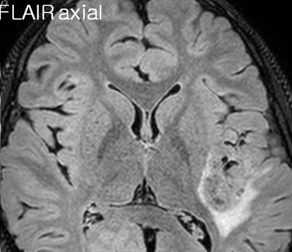
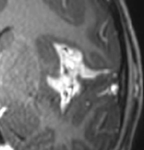
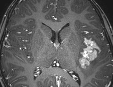
| Discussion | Brain, left (frozen and permanent), removal Meningioangiomatosis Rare, benign hamartomatous cortical/leptomeningeal malformation Growth of meningothelial cells occupying the leptomeninges and involving the cerebral cortex with the formation of a cortical plaque Cortex shows sheathing of the perforating vessels by meningothelial cells, and reactive gliosis Pathogenesis Uncertain uncharacterized vascular malformation direct invasion of a leptomeningeal meningioma into the brain hamartomatous lesion of the leptomeninges and cerebral cortex Single >> multifocal, diffuse May associated NF-2 25% of cases Asymptomatic, multifocal Clinical manifestation Headache, seizure (80%), asymptomatic Refractory and progressive seizure Male > female Children and young adults Location Fronto-parieto-temporal Temporal lobe : single most frequent location (40%) Reports MA with other lesions including meningiomas, hemorrhage, oligodendroglioma, encephalomalacia, AVM Imaging findings CT: Calcified, enhancing lesion with surrounding low density Cortical mass with 80-90% has calcification MR Most commonly hypointense on T1 and hyperintense T2 with about 80% of lesions demonstrating at least some level of enhancement on T1 CE Susceptability artifact Gyriform, can associated cystic component Gyral pattern is maintained but the brain surface may appear abnormal The dense lesion thickens and hardens the affected cortex over a sharply defined plaque-like area, with serpentine vessels on the surface, often creating the impression of a vascular malformation Relatively little mass effect and edema Conventional angiography Normal >> rarely hypervascularity, an abnormal vessel or an avascular mass can be seen References: Meningioangiomatosis: advanced imaging and pathological study of two cases Neuroradiology (1995) 37:120-123 Sporadic meningioangiomatosis-associated atypical meningioma mimicking parenchymal invasion of brain: a case report and review of the literature Chen et al. Diagnostic Pathology 2010, 5:39 Meningioangiomatosis A comprehensive analysis of clinical and laboratory features Brain (1999), 122, 709–726 Cerebral Meningioangiomatosis J Med Sci 2008;28(6):255-258 Meningioangiomatosis without neurofibromatosis simulating encephalitis in neuroimaging Surg Neurol Int 2012;3:34 |
| Correct answer |
Correct Answer
Semi-Correct Answer |