HOME > Weekly Case > Weekly Case
Weekly Case
종결
Case No. 309
2013-07-01~2013-07-31
출제자 : 이영흔
Hit : 352
| Period | 2013-07-01~2013-07-31 |
|---|---|
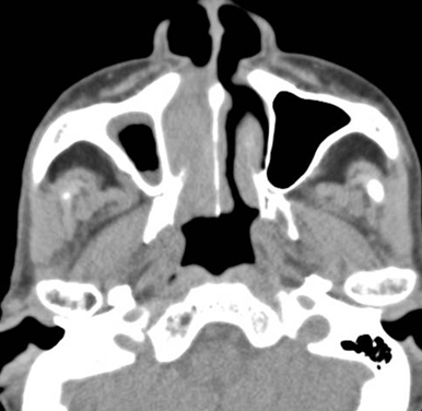
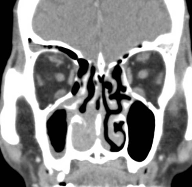
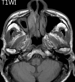
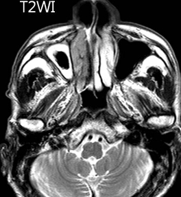
| Diagnosis | Melanoma |
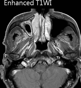
| Clinical information | M/60, RT. nasal cavity mass, 진단은? |
| Discussion | Melanoma Imaging findings: Computed Tomography Soft tissue density Diffuse enhancement Bone destruction with/without remodeling MRI T1WI : high SI due to melanin, best diagnostic clue Diffuse enhancement T2 low SI References Ou, Chang-Hsien et al. Nasal NK/T-cell Lymphoma: Computed Tomography and Magnetic Resonance Imaging Findings. J Clin Med Assoc 2007; 70: 207-212 Kim, Jinna et al. Extranodal nasal-type NK/T-cell lymphoma: Computed tomography findings of head and neck involvement. Acta Radiologica 2010;51:164-169 MR Imaging of Nasal T-Cell/Natural Killer Cell Lymphoma. AJR 2000; 174: 209-211 Xie, Chuan-miao et al. Primary nasopharyngeal non-Hodgkin’s lymphoma: imaging patterns on MR imaging. Clinical Imaging 2013; 37: 458-464 |
| Correct answer |
Correct Answer
Semi-Correct Answer |