HOME > Weekly Case > Weekly Case
Weekly Case
종결
Case No. 307
2013-06-03~2013-06-30
출제자 : 안성준
Hit : 453
| Period | 2013-06-03~2013-06-30 |
|---|---|
| Diagnosis | progressive necrotizing malignant external otitis |
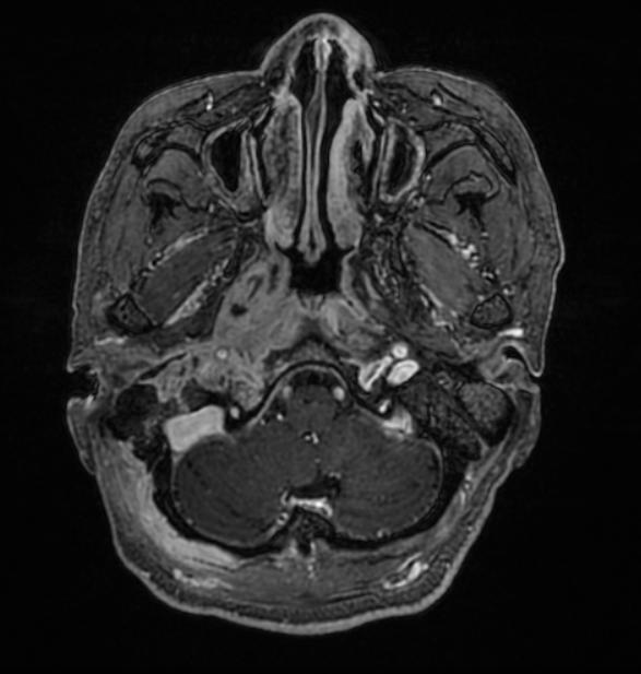
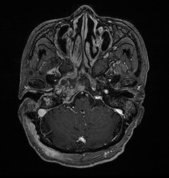
| Clinical information | 64/F, C.C : Headache for severeal months, Recently aggravation, facial palsy 아래 3개 영상 --> 3개월 후 추적 MRI |
| Discussion | progressive necrotizing malignant external otitis 사진해설> 3개월 전 MRI 는 Rt, temporal bone과 ECA 주위로 enhancement가 있어 Post op granulation 으로 생각할 수 있으나 최근 MRI 를 보게되면 inflammation 이 Rt. Nasopharynx와 retropharyngeal space, skull base, Rt. Posterior deep neck까지 퍼진 것을 관찰 할 수 있으며 환자가 DM의 history를 가지고 있는 것을 고려할 때 necotizing malignant external otitis로 진단 할 수 있다. Review> Necrotizing (malignant) external otitis, an infection involving the temporal and adjacent bones, is a relatively rare complication of external otitis. It occurs primarily in immunocompromised persons, especially older persons with diabetes mellitus, and is often initiated by self-inflicted or iatrogenic trauma to the external auditory canal. The most frequent pathogen is Pseudomonas aeruginosa. Clinical findings include granulation tissue in the external auditory canal, especially at the bone-cartilage junction. Facial and other cranial nerve palsies indicate a poor prognosis; intracranial complications are the most frequent cause of death[1] Reference 1. Parisier SC, Lucente FE, Som PM, Hirschman SZ, Arnold LM, et al. (1982) Nuclear scanning in necrotizing progressive "malignant" external otitis. Laryngoscope 92: 1016-1019. |
| Correct answer |
Correct Answer
Semi-Correct Answer |